Gallery
RIED
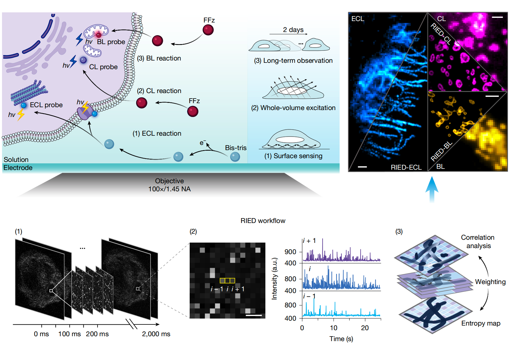
System:
Reaction-luminescence super-resolution imaging.
Camera:
EMCCD camera (iXon Ultra 897, Andor).
Description:
RIED combines entropy-weighted correlation with deconvolution to reconstruct super-resolution ECL, CL, and BL images at approximately 100 nm resolution, without external light excitation.
RIED / ECLConventional ECL ↔ RIED reconstruction
System:
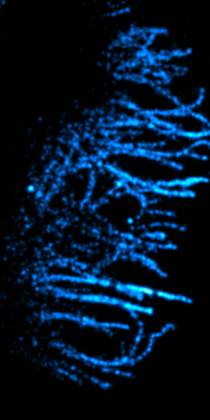
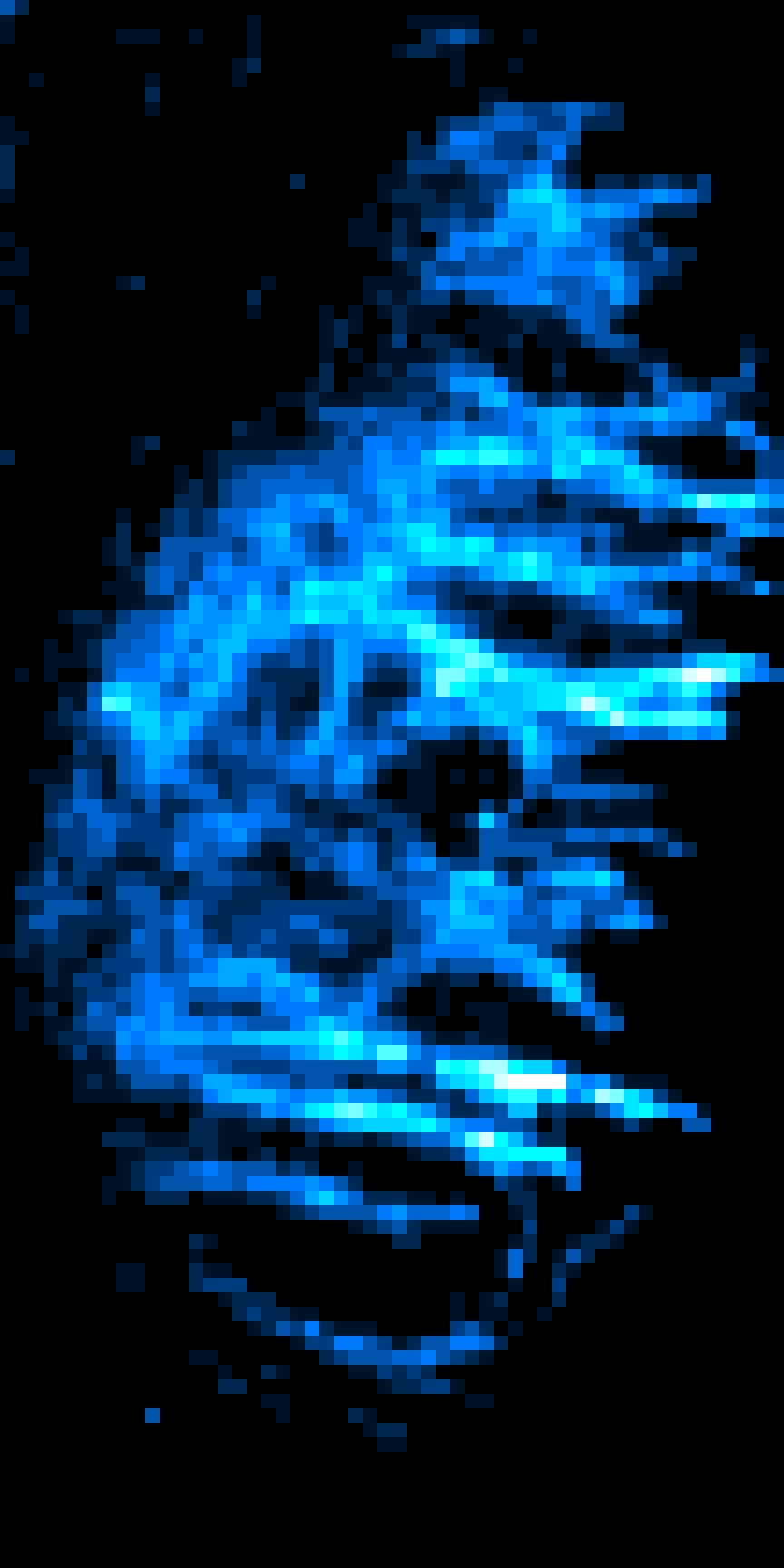
Electrochemiluminescence super-resolution imaging (ECL-RIED).
Objective:
Oil-immersion 100× objective (NA = 1.45, Olympus).
Camera:
EMCCD camera (iXon Ultra 897, Andor).
Sample:
Microtubules in a HeLa cell.
Label:
α-tubulin immunostaining with Ru(bpy)32+-conjugated antibodies.
Description:
Left: conventional ECL; Right: ECL-RIED, reaching 97 nm resolution.
RIED / CLConventional CL ↔ RIED reconstruction
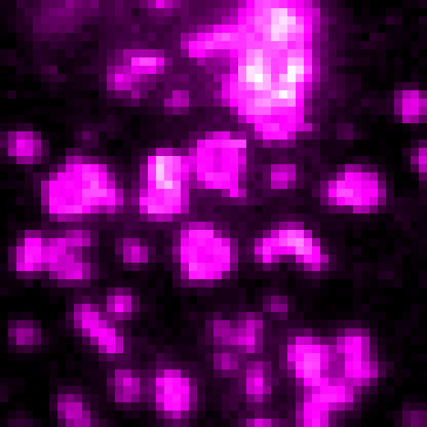
System:
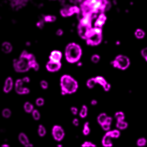
Chemiluminescence super-resolution imaging (CL-RIED).
Objective:
Oil-immersion 100× objective (NA = 1.45, Olympus).
Camera:
EMCCD camera (iXon Ultra 897, Andor).
Sample:
Mitochondria in fixed COS-7 cells.
Label:
Mitochondria-targeted GeNL (NanoLuc–mNeonGreen BRET probe).
Description:
Left: conventional CL; Right: CL-RIED, achieving 102 nm lateral and 221 nm axial resolution for three-dimensional mitochondrial mapping.
RIED / BLConventional BL ↔ RIED reconstruction
System:
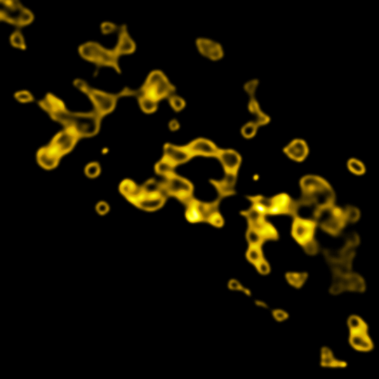
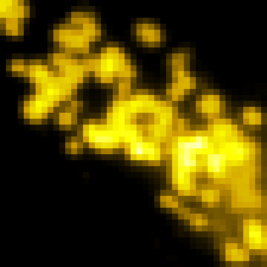
Bioluminescence super-resolution imaging (BL-RIED).
Objective:
Oil-immersion objectives (100×/1.42 NA or 60×/1.49 NA, Nikon).
Camera:
EMCCD camera (iXon Ultra 897, Andor).
Sample:
Mitochondrial outer membranes in live COS-7 cells.
Label:
Mitochondrial-outer-membrane-targeted GeNL (NanoLuc–mNeonGreen BRET probe).
Description:
Left: conventional BL; Right: BL-RIED. It reaches 117 nm resolution and supports continuous live-cell imaging for 41 h.
RIED / 2D ECLConventional 2D ECL ↔ RIED reconstruction
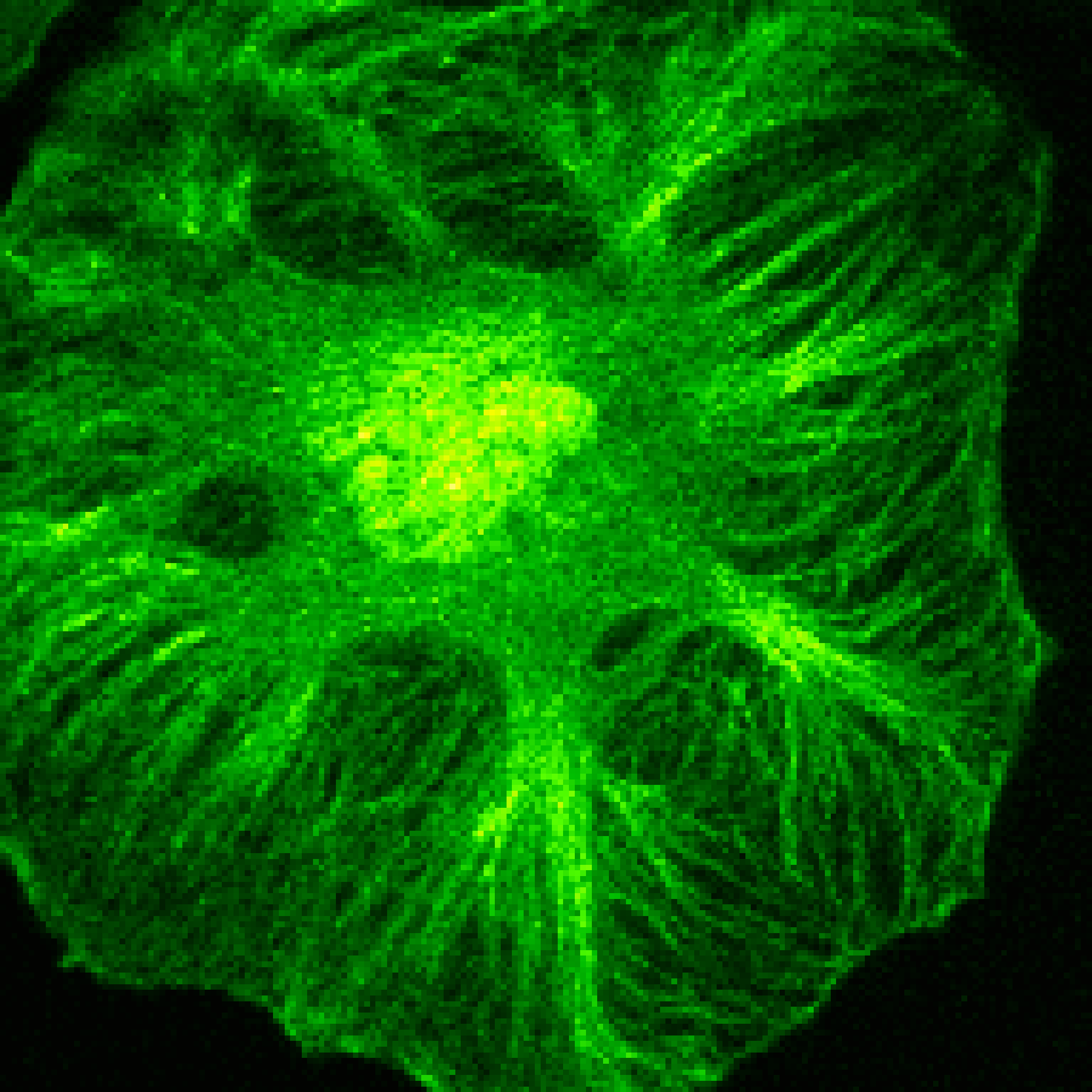
System:
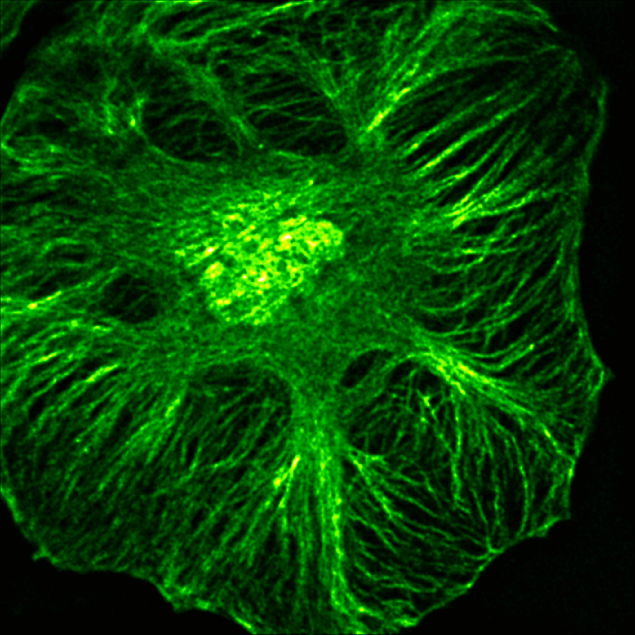
Two-dimensional ECL-RIED.
Objective:
Oil-immersion 100× objective (NA = 1.45, Olympus).
Camera:
EMCCD camera (iXon Ultra 897, Andor).
Sample:
Microtubules in a HeLa cell.
Label:
α-tubulin immunostaining with Ru(bpy)32+-conjugated antibodies.
Description:
Left: conventional 2D ECL; Right: 2D ECL-RIED with substantially improved structural detail.
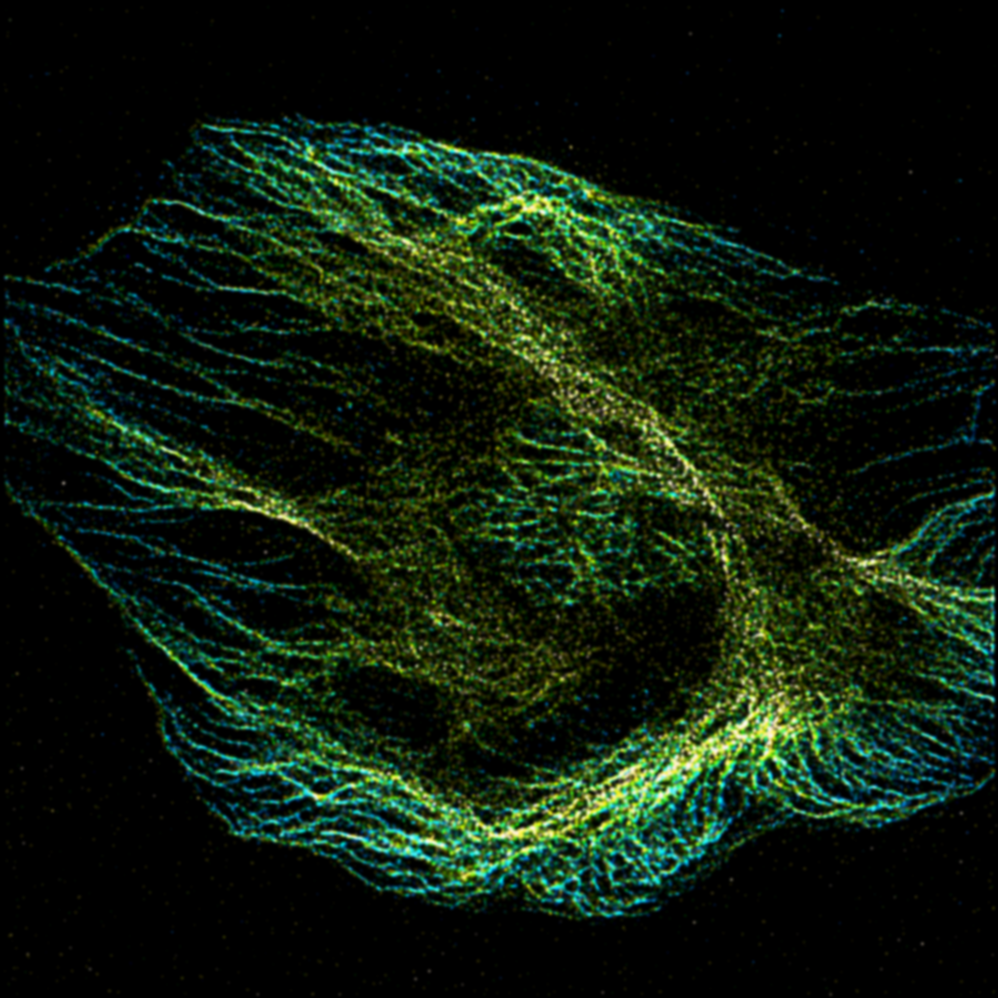
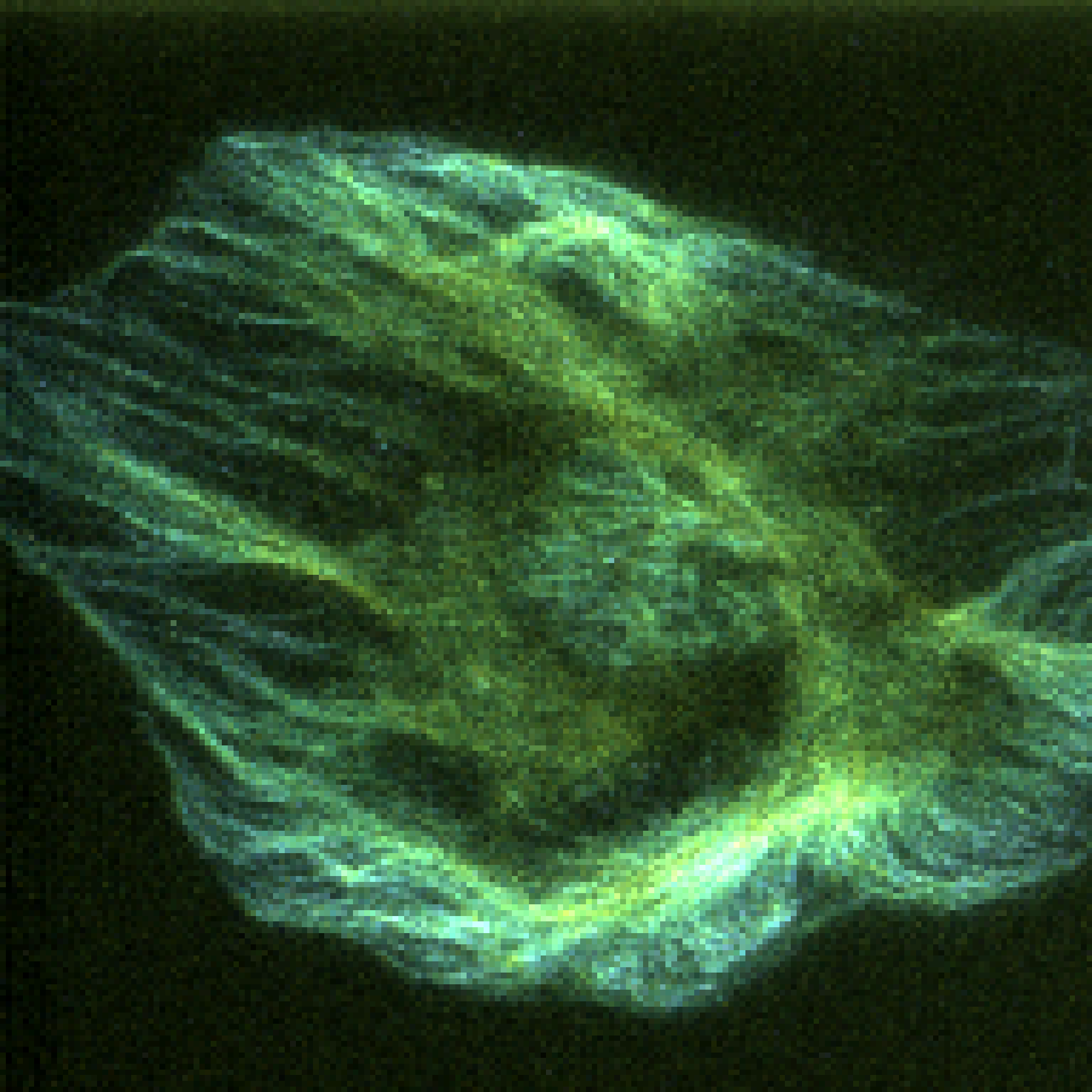
RIED / 3D ECLConventional 3D ECL ↔ RIED reconstruction
System:
Three-dimensional ECL-RIED.
Objective:
Oil-immersion 100× objective (NA = 1.45, Olympus).
Camera:
EMCCD camera (iXon Ultra 897, Andor).
Sample:
Microtubules in a HeLa cell.
Label:
α-tubulin immunostaining with Ru(bpy)32+-conjugated antibodies.
Color code:
Color represents axial depth, distinguishing structures at different z positions.
Description:
Left: conventional 3D ECL; Right: 3D ECL-RIED, resolving approximately 116 nm laterally and 235 nm axially.
SN2N
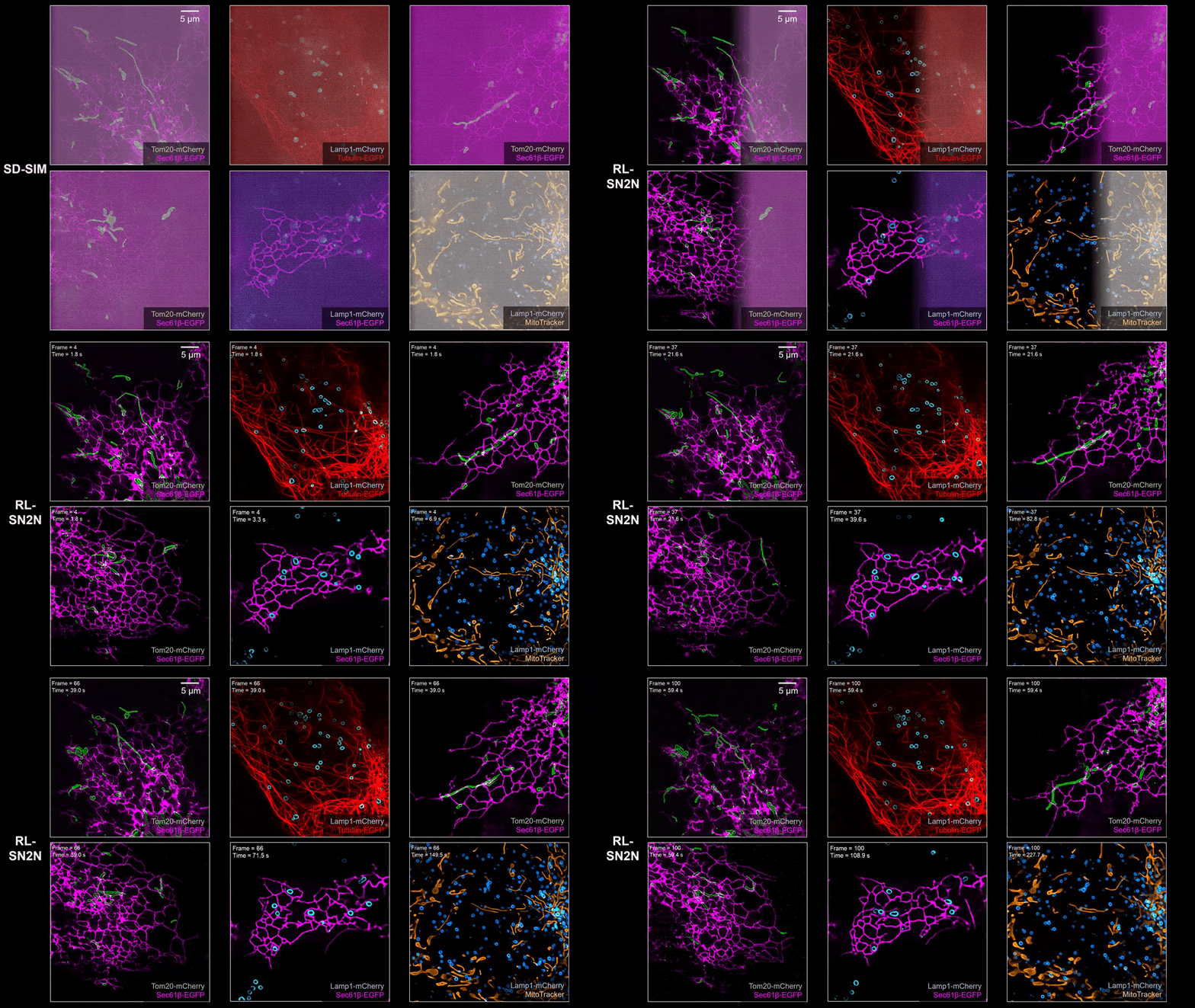
SN2N / SD-SIMNoisy acquisition ↔ SN2N denoising
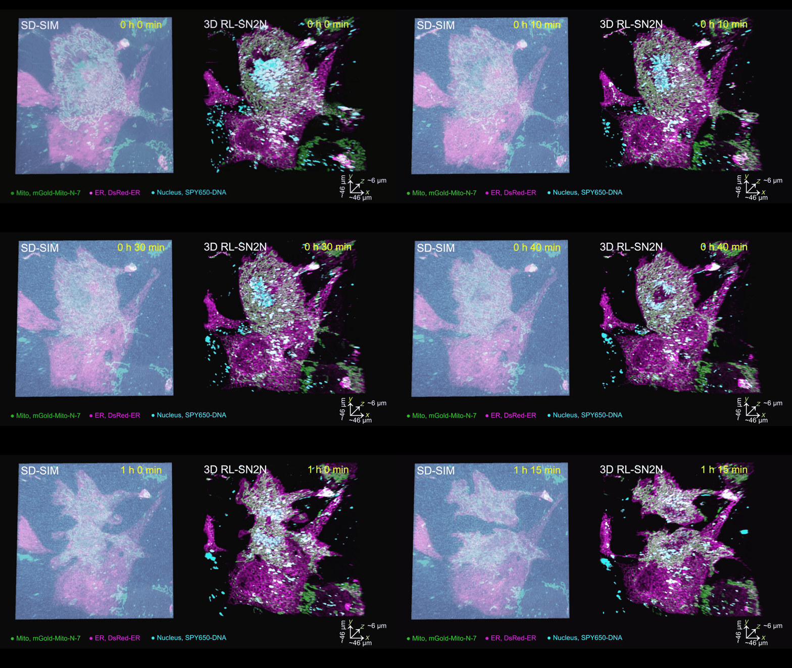
System:
SD-SIM.
Objective:
Wide-field objective (100×/1.3 oil, Olympus)
Camera:
sCMOS camera (C14440-20UP, Hamamatsu, Japan).
Cell line:
COS-7 cell.
Description:
RL-SN2N on SD-SIM.
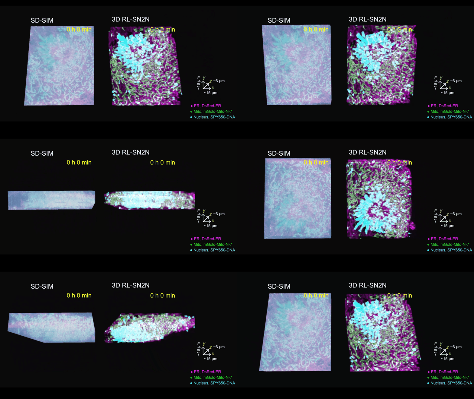
System:
SD-SIM.
Objective:
Wide-field objective (100×/1.3 oil, Olympus)
Camera:
sCMOS camera (C14440-20UP, Hamamatsu, Japan).
Cell line:
COS-7 cell.
Description:
3D RL-SN2N on SD-SIM.
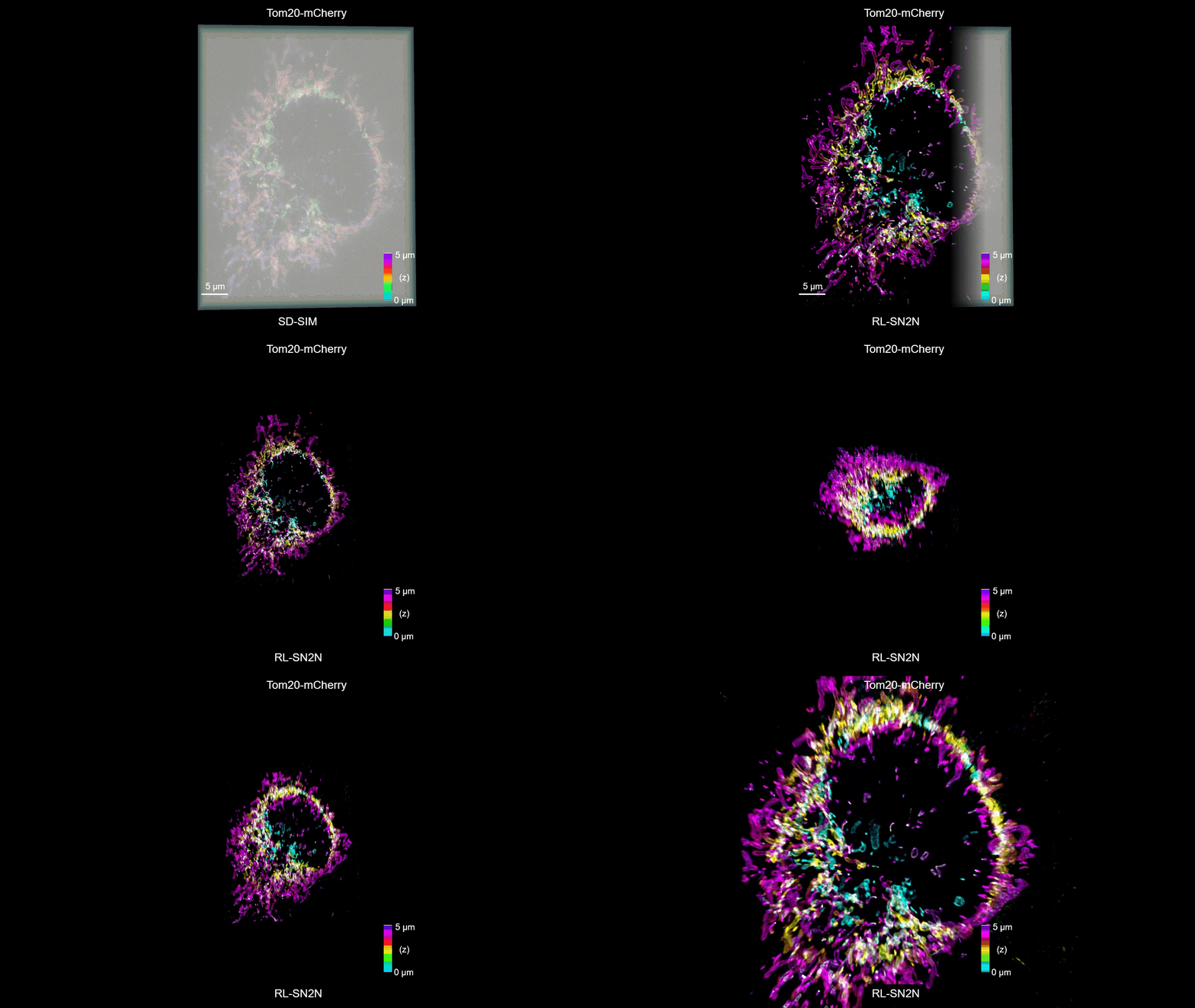
System:
SD-SIM.
Objective:
Wide-field objective (100×/1.3 oil, Olympus)
Camera:
sCMOS camera (C14440-20UP, Hamamatsu, Japan).
Cell line:
COS-7 cell.
Description:
3D RL-SN2N on SD-SIM.
System:
SD-SIM.
Objective:
Wide-field objective (100×/1.3 oil, Olympus)
Camera:
sCMOS camera (C14440-20UP, Hamamatsu, Japan).
Cell line:
COS-7 cell.
Description:
3D RL-SN2N on SD-SIM.
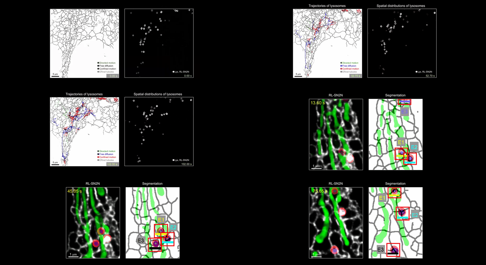
System:
SD-SIM.
Objective:
Wide-field objective (100×/1.3 oil, Olympus)
Camera:
sCMOS camera (C14440-20UP, Hamamatsu, Japan).
Cell line:
COS-7 cell.
Description:
RL-SN2N on SD-SIM.
SN2N / STEDNoisy acquisition ↔ SN2N denoising
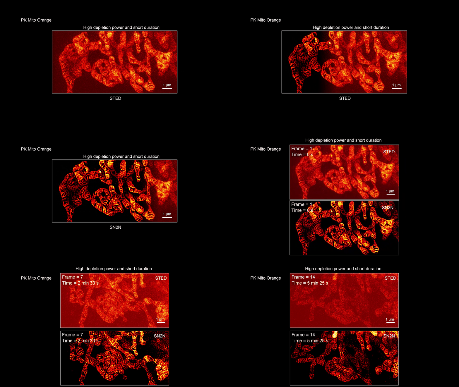
System:
STED.
Objective:
Wide-field objective (100×/1.49 oil, Olympus) Cell line:
COS-7 cell.
Description:
SN2N on STED.
SN2N / GI-SIMNoisy acquisition ↔ SN2N denoising
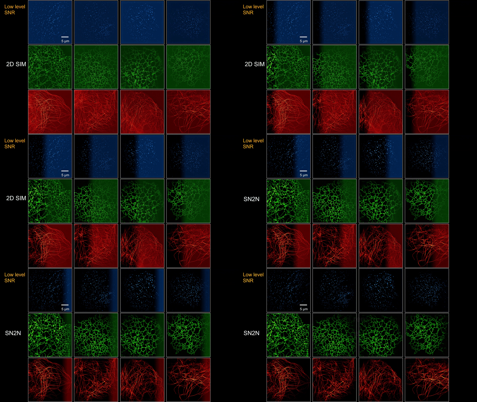
SN2N / 2D-SIMNoisy acquisition ↔ SN2N denoising
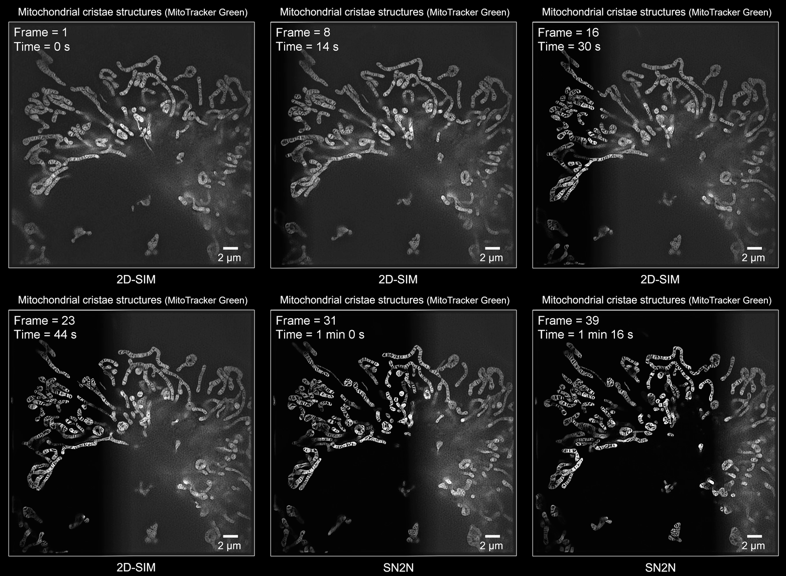
System:
2D-SIM.
Objective:
Wide-field objective (100×/1.49 oil, Olympus) Cell line:
COS-7 cell.
Camera:
sCMOS camera (C14440-20UP, Hamamatsu, Japan).
Description:
SN2N on SIM.
Sparse deconvolution-assisted microscopy
Sparse deconvolution / SD-SIMRaw acquisition ↔ Sparse reconstruction
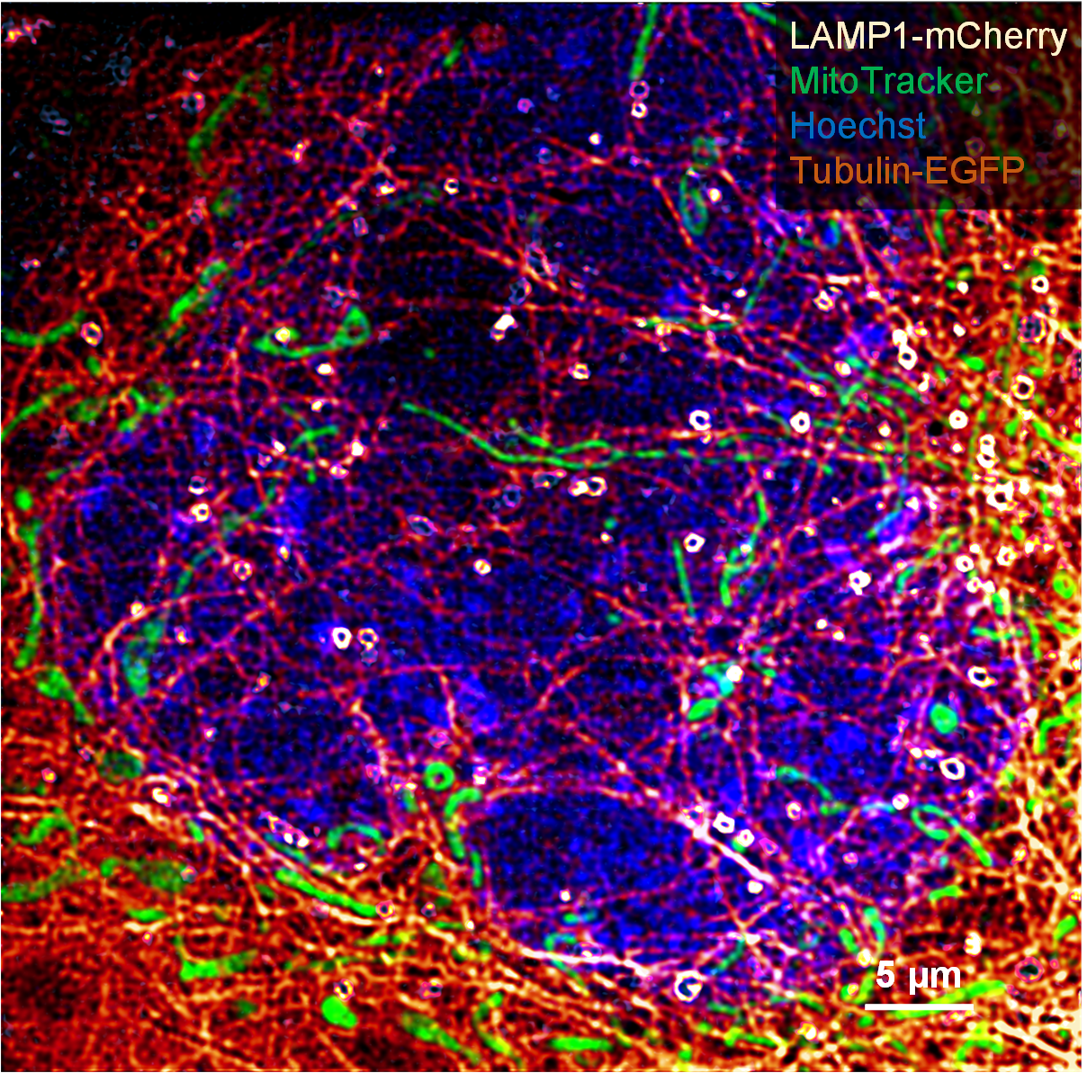
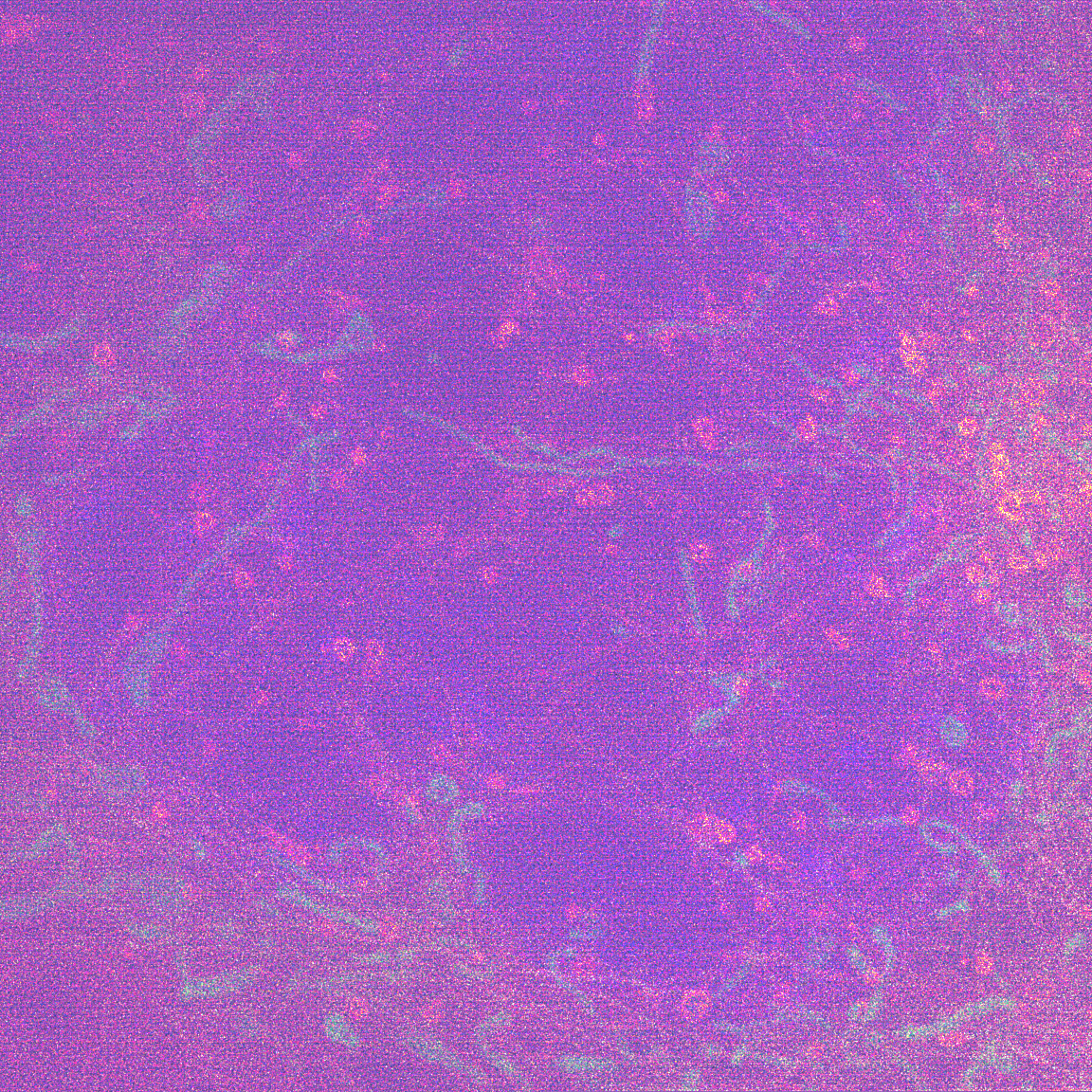
System:
SD-SIM.
Objective:
Wide-field objective (100×/1.3 oil, Olympus)
Camera:
sCMOS camera (C14440-20UP, Hamamatsu, Japan).
Cell line:
COS-7 cell.
Label:
Lamp1-mCherry;
MitoTracker® Deep Red FM;
Hoechst H1399;
Tubulin-EGFP.
Description:
Left: raw SD-SIM; Right: Sparse SD-SIM.
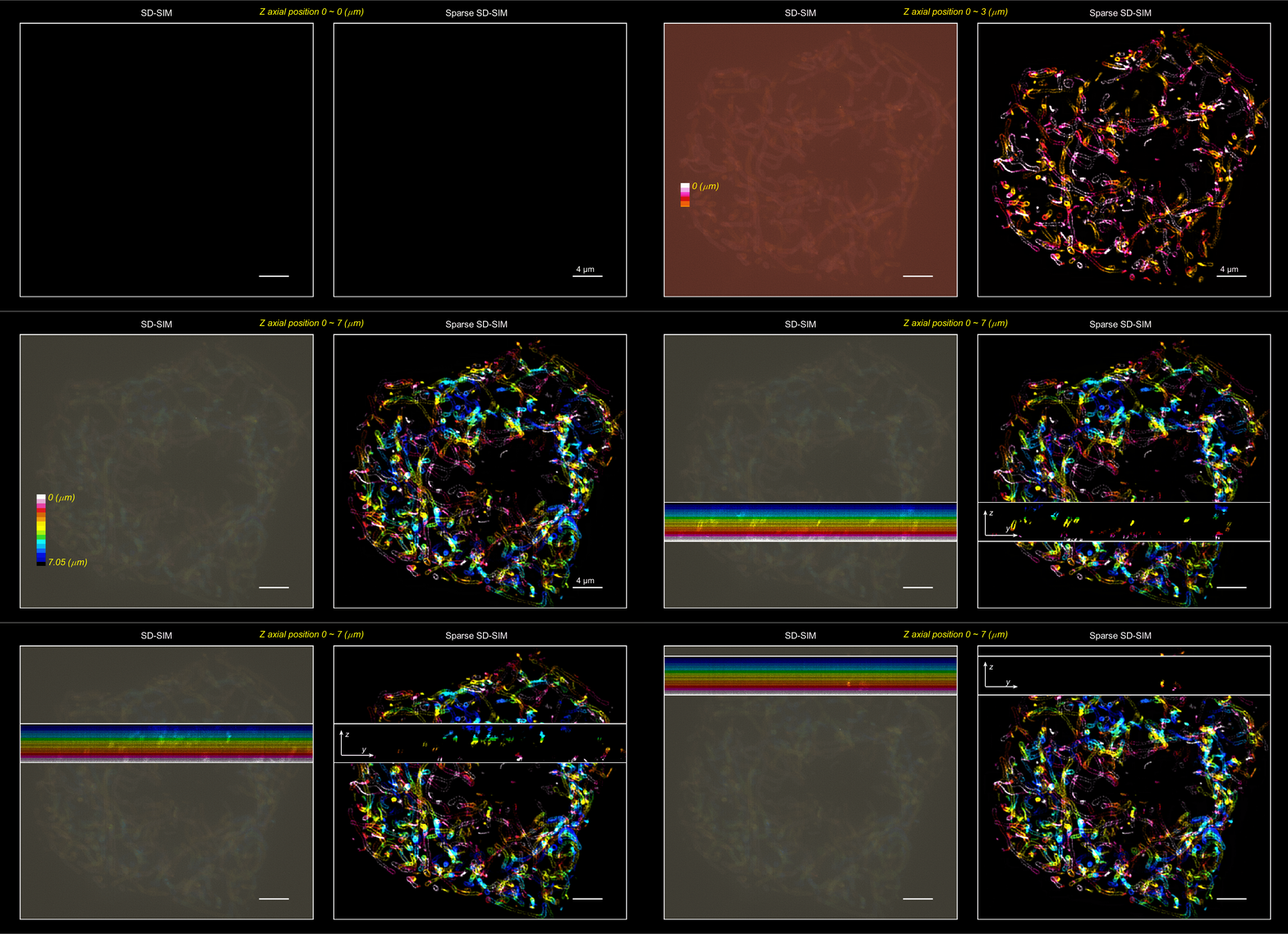
System:
SD-SIM.
Objective:
Wide-field objective (100×/1.3 oil, Olympus)
Camera:
sCMOS camera (C14440-20UP, Hamamatsu, Japan).
Cell line:
COS-7 cell.
Label:
Tom20-mCherry;
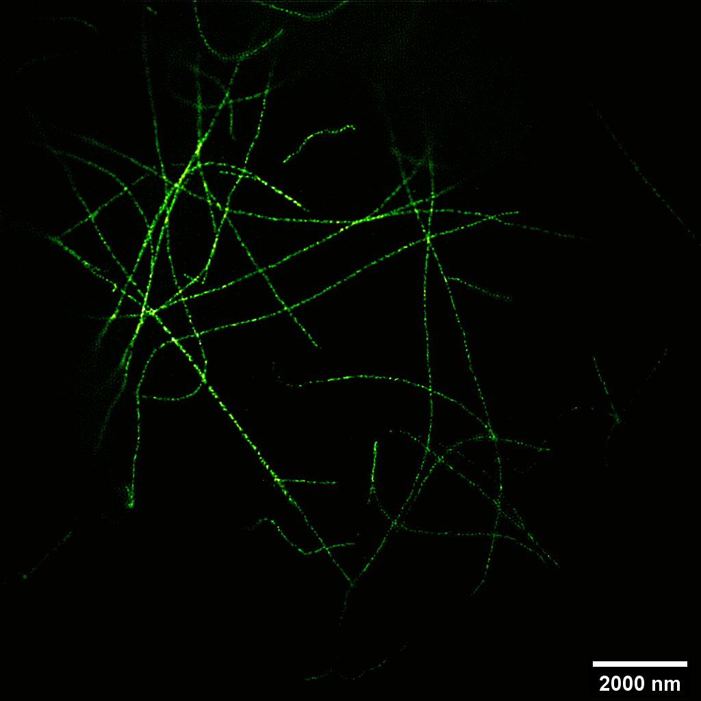
Sparse deconvolution / 4.5× expansion microscopyRaw expansion ↔ Sparse reconstruction
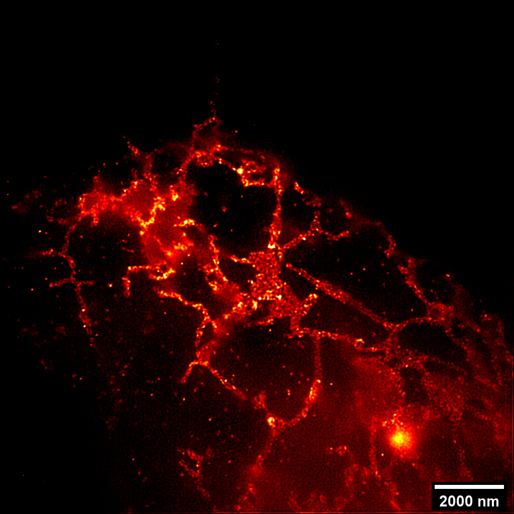
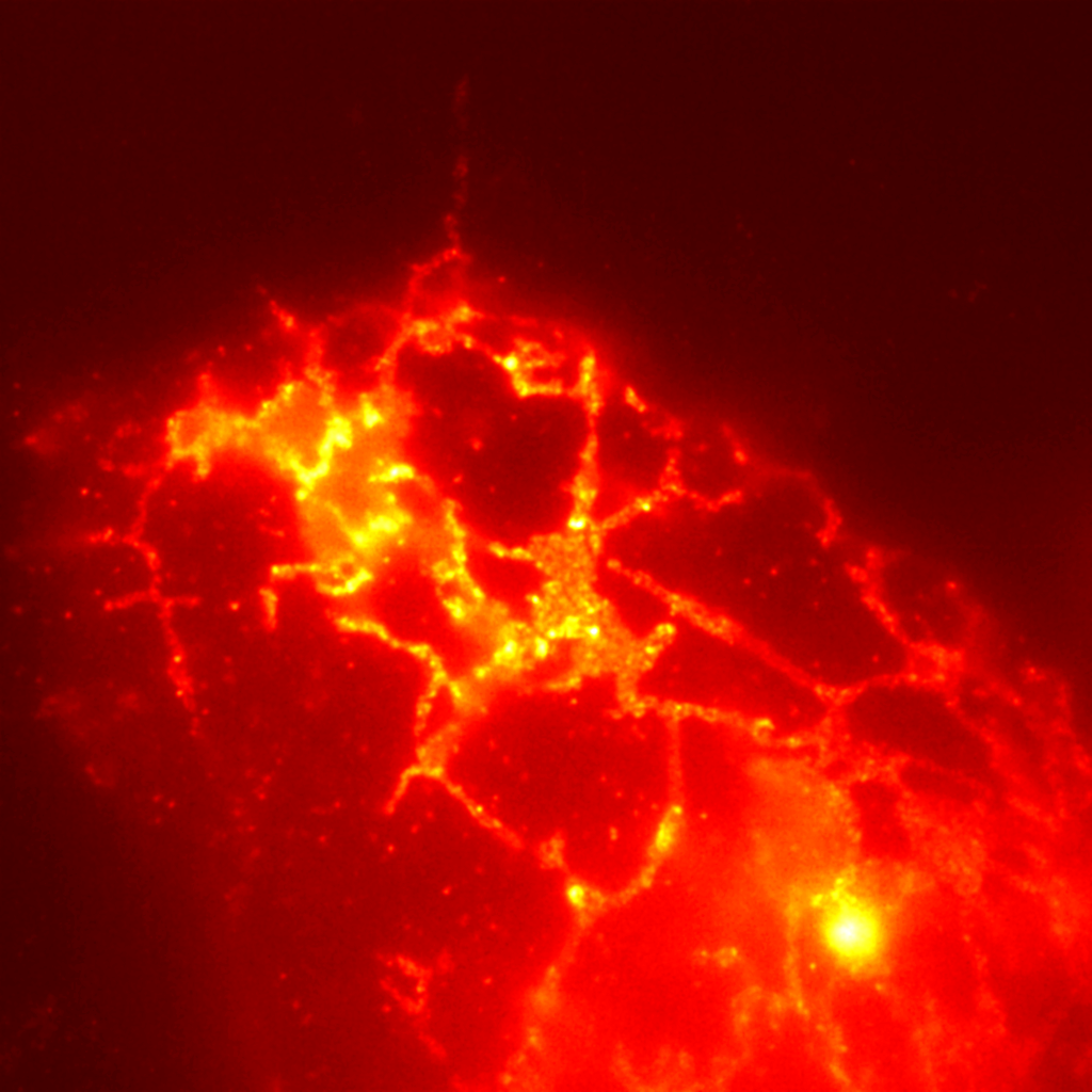
System:
Expansion microscopy 4.5-fold.
Objective:
Wide-field objective (100×/1.45 oil, Olympus)
Camera:
sCMOS camera (Flash 4.0 V3, Hamamatsu, Japan)
Cell line:
COS-7 cell.
Label:
Sec61β-GFP.
Description:
Left: raw expansion; Right: Sparse expansion.
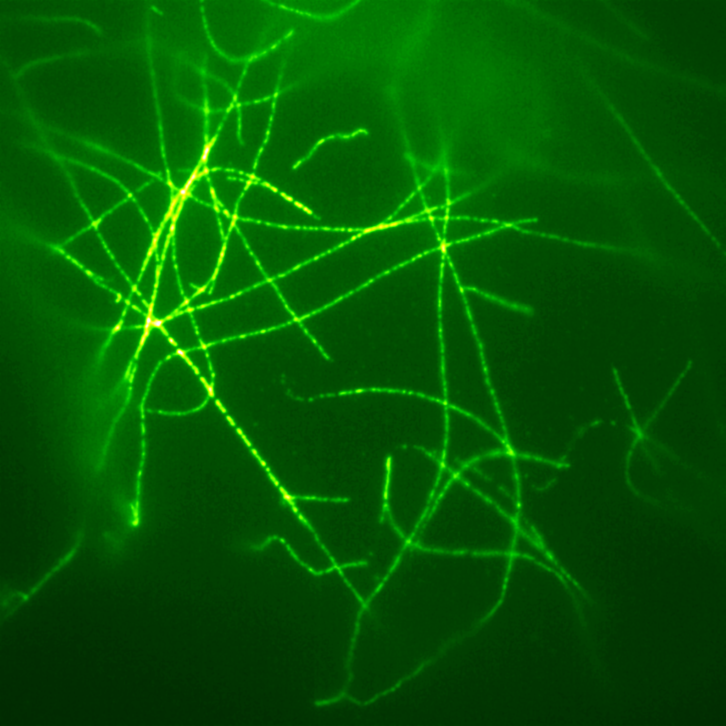
System:
Expansion microscopy 4.5-fold.
Objective:
Wide-field objective (100×/1.45 oil, Olympus)
Camera:
sCMOS camera (Flash 4.0 V3, Hamamatsu, Japan)
Cell line:
COS-7 cell.
Label:
α-tubulin immunostaining.
Description:
Left: raw expansion; Right: Sparse expansion.
Sparse deconvolution / 2D-SIMRaw SIM ↔ Sparse reconstruction
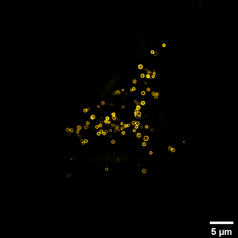
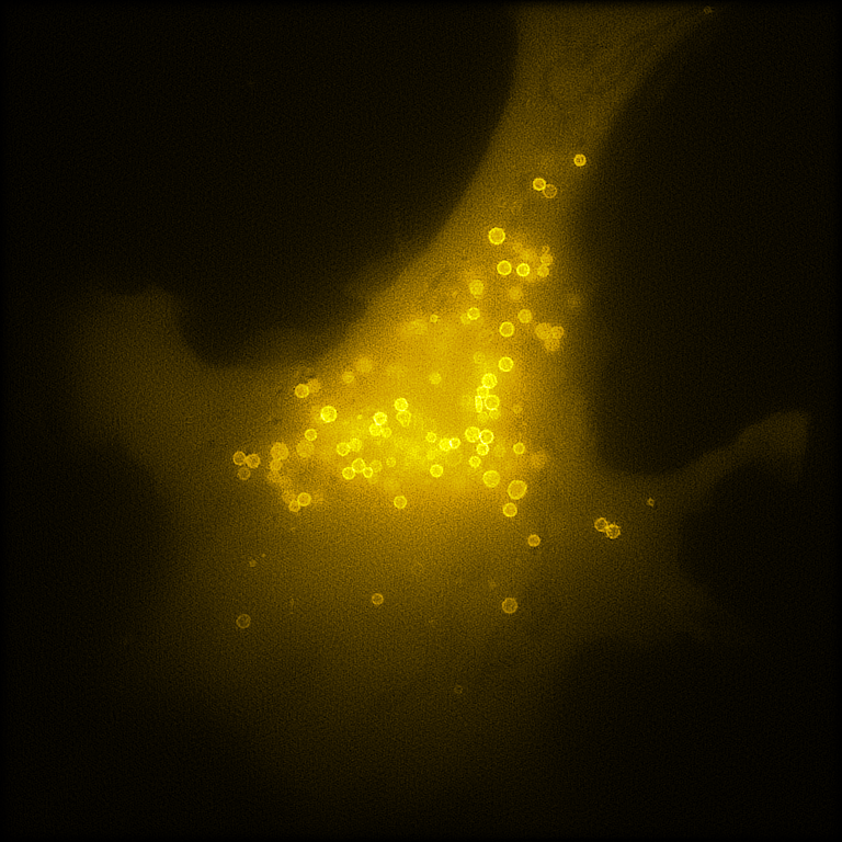
System:
2D-SIM.
Objective:
TIRF objective (100×/1.49 oil, Olympus)
Camera:
sCMOS camera (Flash 4.0 V3, Hamamatsu, Japan)
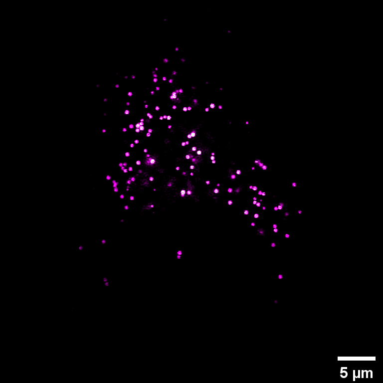
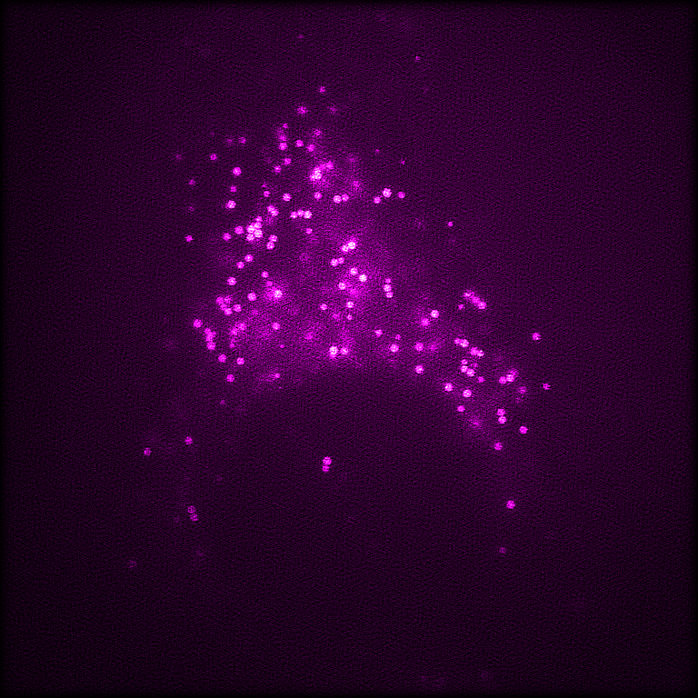
Cell line:
COS-7 cell.
Label:
LAMP1-EGFP.
Description:
Left: raw 2D-SIM; Right: Sparse 2D-SIM.
System:
2D-SIM.
Objective:
TIRF objective (100×/1.49 oil, Olympus)
Camera:
sCMOS camera (Flash 4.0 V3, Hamamatsu, Japan)
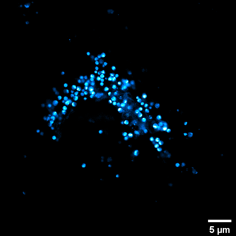
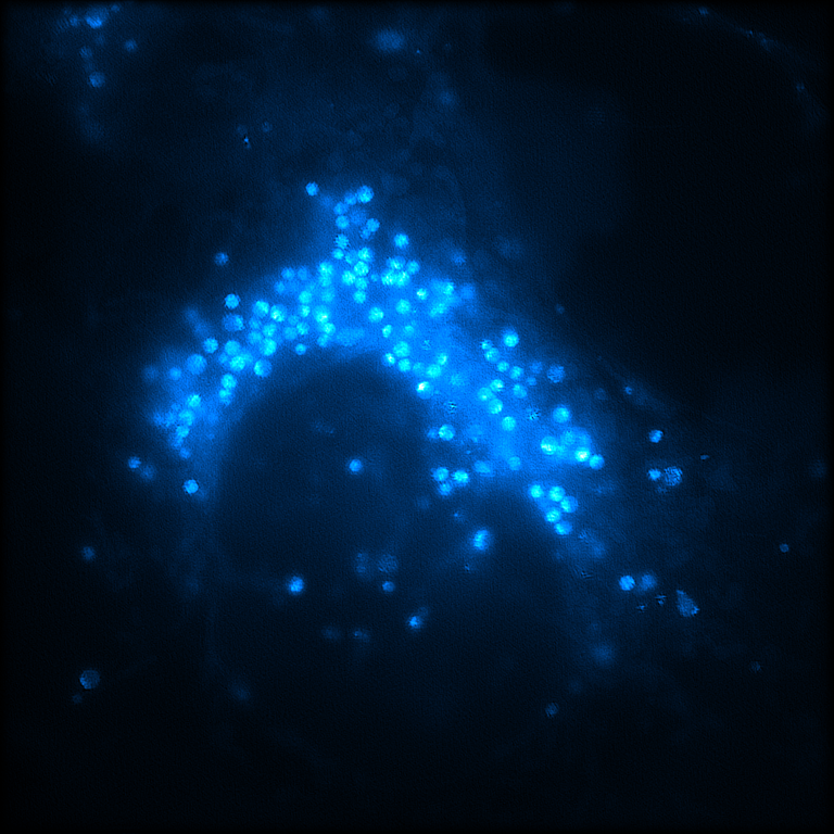
Cell line:
COS-7 cell.
Label:
LipidSpot 488.
Description:
Left: raw 2D-SIM; Right: Sparse 2D-SIM.
System:
2D-SIM.
Objective:
TIRF objective (100×/1.49 oil, Olympus)
Camera:
sCMOS camera (Flash 4.0 V3, Hamamatsu, Japan)
Cell line:
COS-7 cell.
Label:
LysoView 488.
Description:
Left: raw 2D-SIM; Right: Sparse 2D-SIM.
Sparse deconvolution / TIRF-SIMRaw SIM ↔ Sparse reconstruction
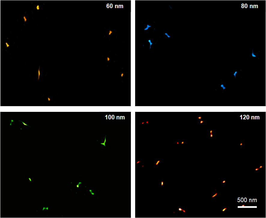
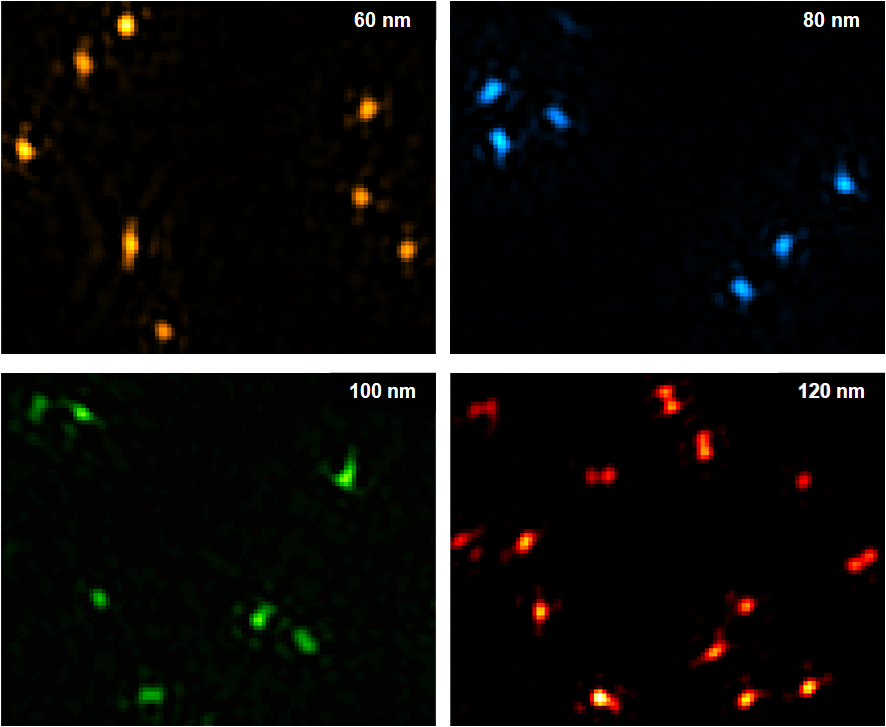
System:
High NA TIRF-SIM.
Objective:
TIRF objective (100×/1.7 HI oil, Olympus)
Camera:
sCMOS camera (Flash 4.0 V3, Hamamatsu, Japan)
Cell line:
60-80-100-120 nm DNA origami.
Label:
Cy5, two sites fluorescently labeled.
Description:
Left: raw 2D-SIM; Right: Sparse 2D-SIM.
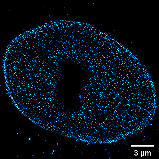
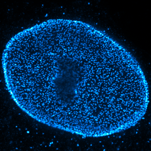
Sparse deconvolution / gated STEDRaw STED ↔ Sparse reconstruction
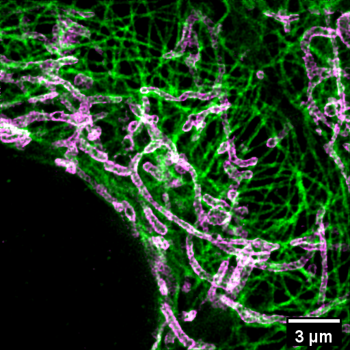
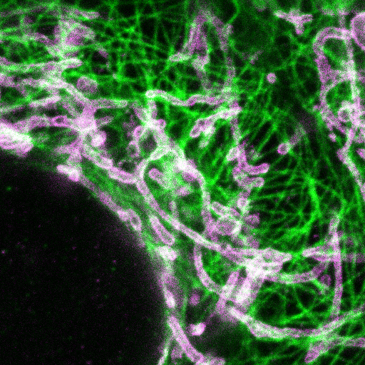
System:
Gated STED microscope.
Objective:
Wide-field objective (100×/1.40 oil, HCX PL APO, Leica)
Cell line:
HeLa cell.
Label:
anti-Tom20;
anti-Tubulin;
Description:
Left: raw STED; Right: Sparse STED.
System:
Gated STED microscope.
Objective:
Wide-field objective (100×/1.40 oil, HCX PL APO, Leica)
Cell line:
HeLa cell.
Label:
anti-Mab414;
Description:
Left: raw STED; Right: Sparse STED.
Sparse deconvolution / MTPMRaw multiphoton image ↔ Sparse reconstruction
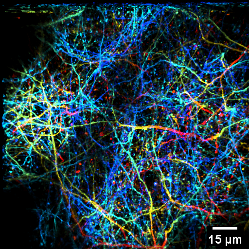
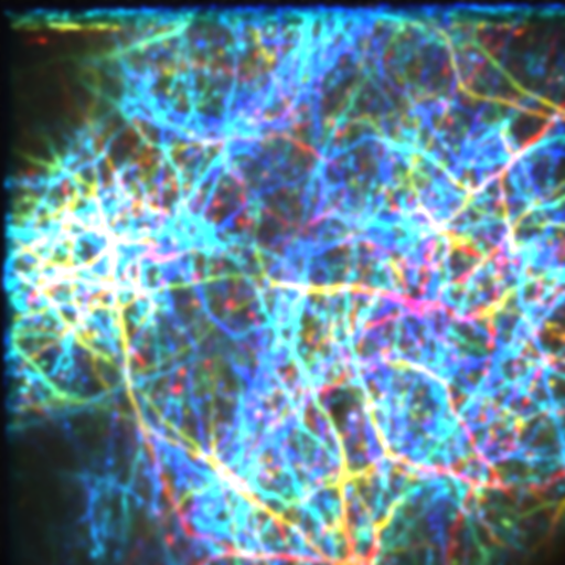
System:
MTPM.
Objective:
Micro-objective with NA 0.7
Sample:
Brain of a transgenic mouse (in vivo).
Label:
Thy1-GFP
Description:
Left: raw MTPM; Right: Sparse MTPM.
Color-coded 3D volume (50~160 μm).
SPoSI
System:
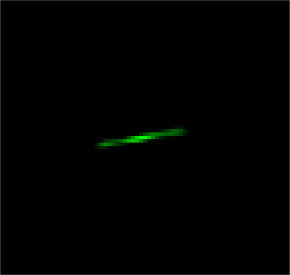
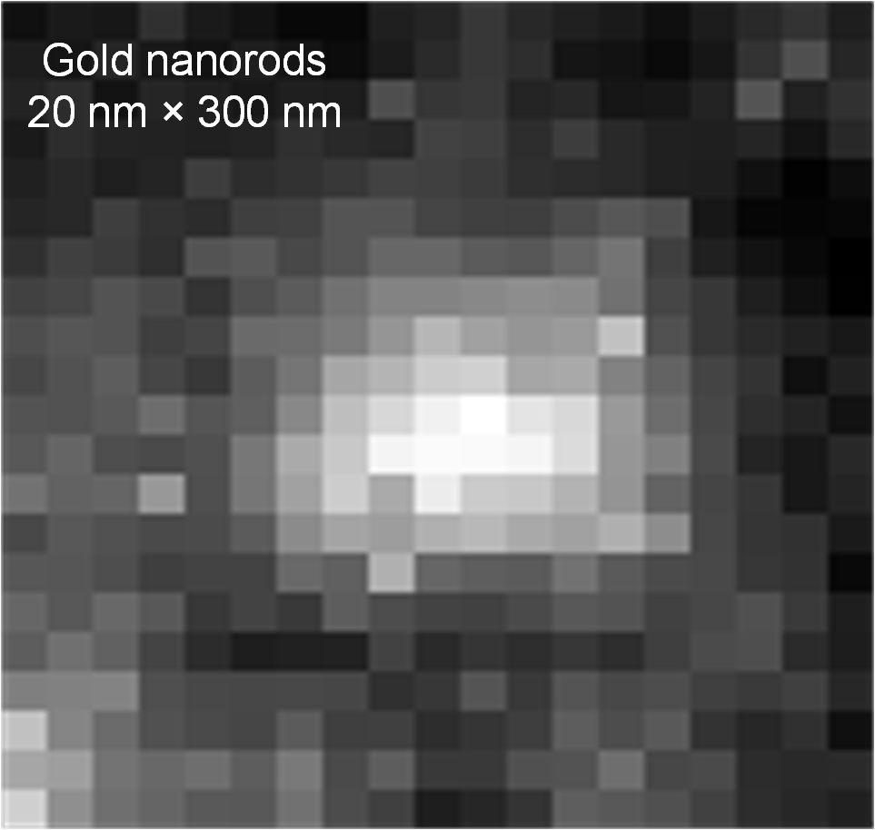
Label-free super-resolution microscopy (SPoSI).
Objective:
Wide-field objective (NA 0.9)
Sample:
Gold nanorod (20 nm x 300 nm).
Description:
Left: raw wide-field (552 nm laser); Right: SPoSI.
Endoscope
System:
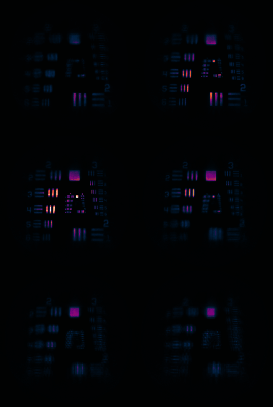
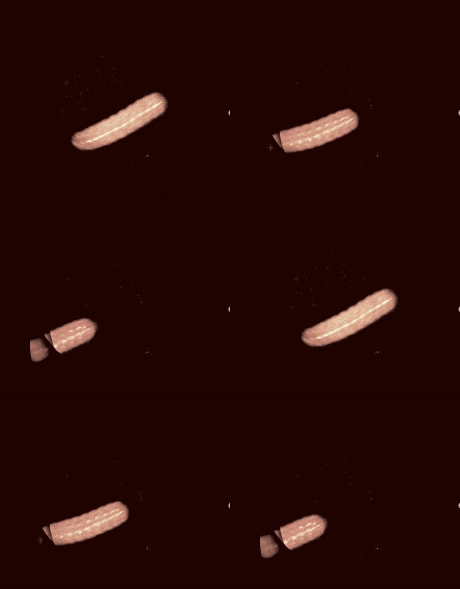
Volumetric endoscope probe.
Objective:
GRIN lens (NA 0.5).
Sample:
USAF target and live worm.
Description:
Left: 3D live worm; Right: tilted USAF target.
Smart palm-size optofluidic hematology analyzer
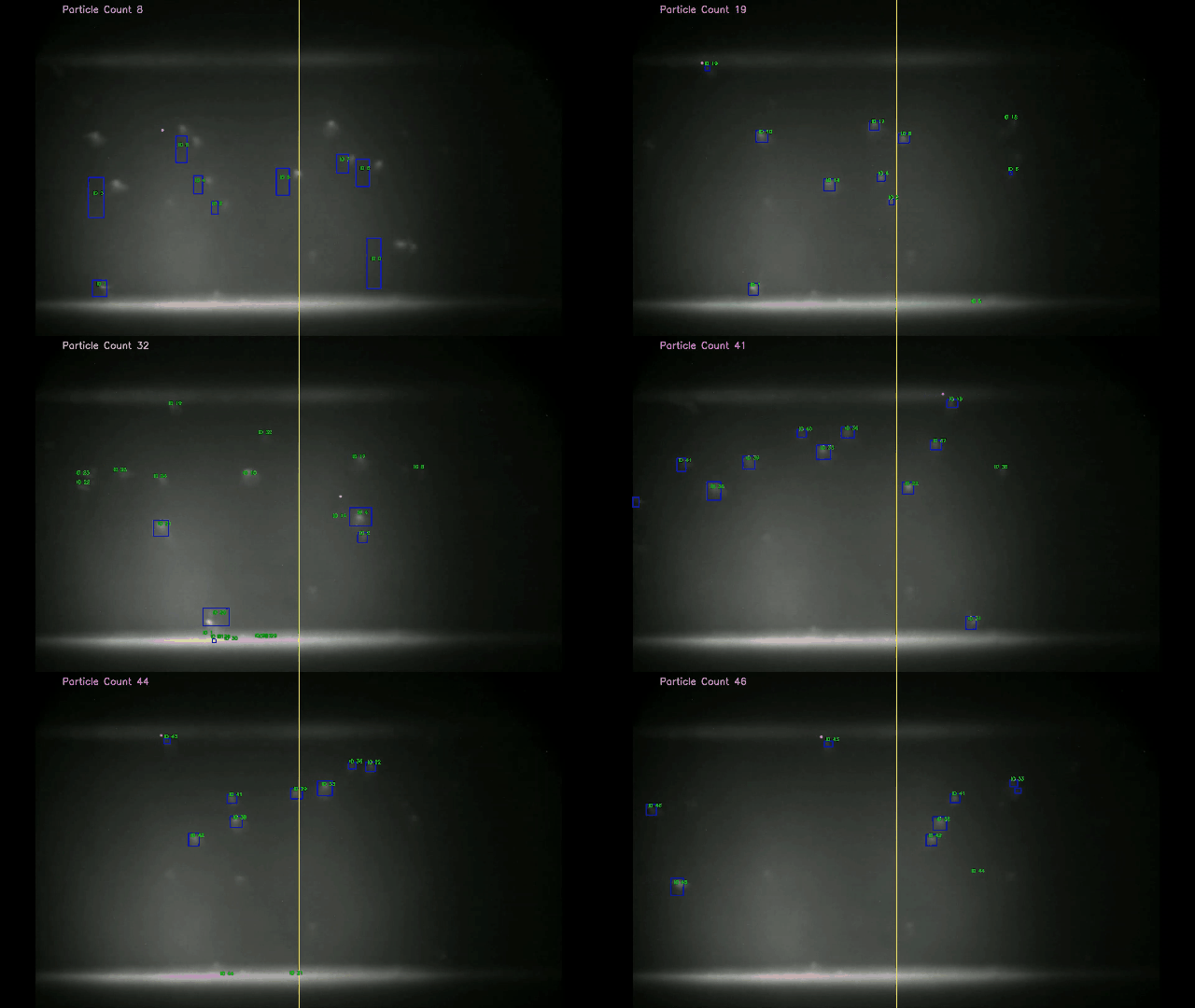
System:
A smart palm-sized optofluidic hematology analyzer based on a miniature fluorescence microscope and a microfluidic platform to reduce the device weight and improve portability.
Objective:
GRIN lens (NA 0.5).
Sample:
Leukocyte.
Description:
Tracking of the leukocytes.
SACD
SACD / spinning-disk confocalConfocal reconstruction ↔ SACD
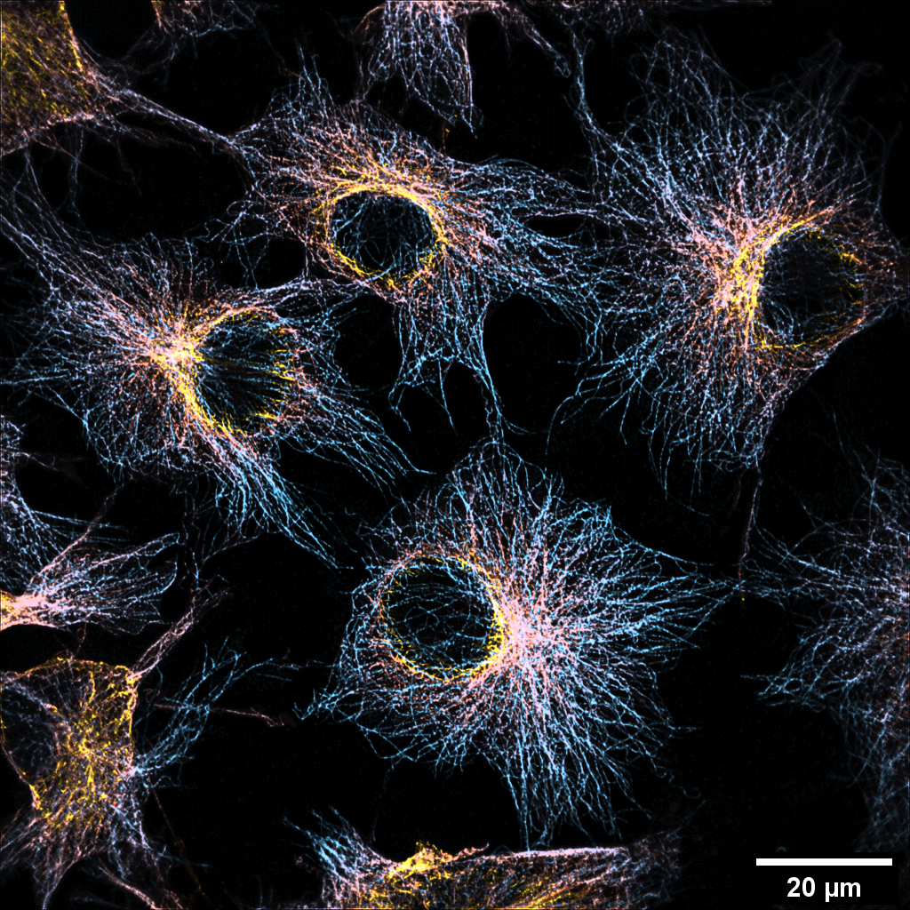
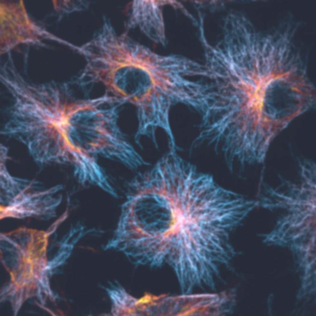
System:
Spinning disk confocal.
Objective:
Wide-field objective (100×/1.3 oil, Olympus)
Camera:
sCMOS camera (Flash 4.0 V3, Hamamatsu, Japan)
Cell line:
COS-7 cell.
Label:
Qdots 525.
Description:
Left: confocal; Right: SACD.
Huge FOV 3D volume (10μm in depth);
20 frame per SACD reconstruction.
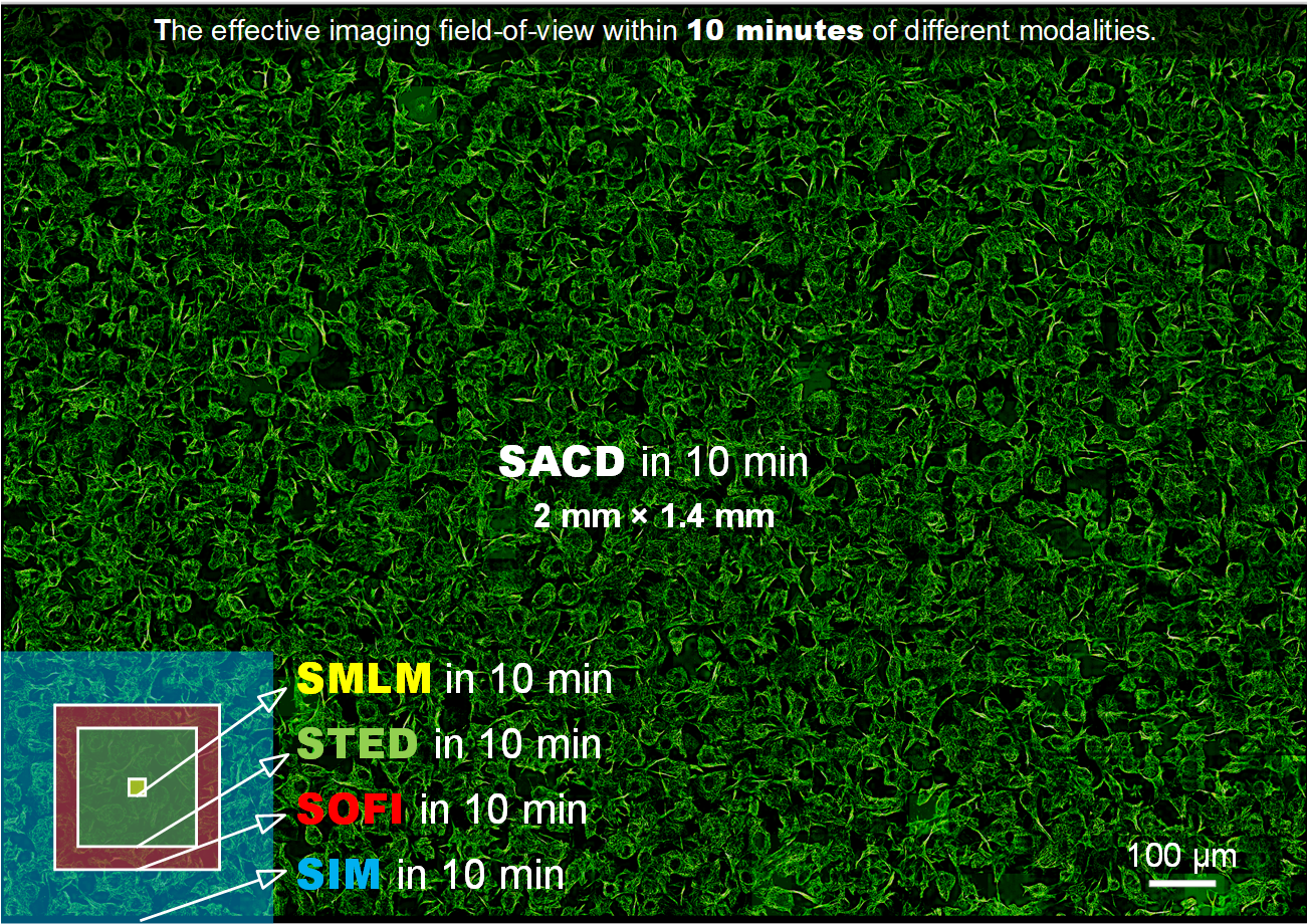
System:
Spinning disk confocal.
Objective:
Wide-field objective (100×/1.3 oil, Olympus)
Camera:
sCMOS camera (Flash 4.0 V3, Hamamatsu, Japan)
Cell line:
COS-7 cell.
Label:
Qdots 525.
20 frame per SACD reconstruction.
PANEL
PANEL / STORMSTORM reconstruction ↔ local resolution map
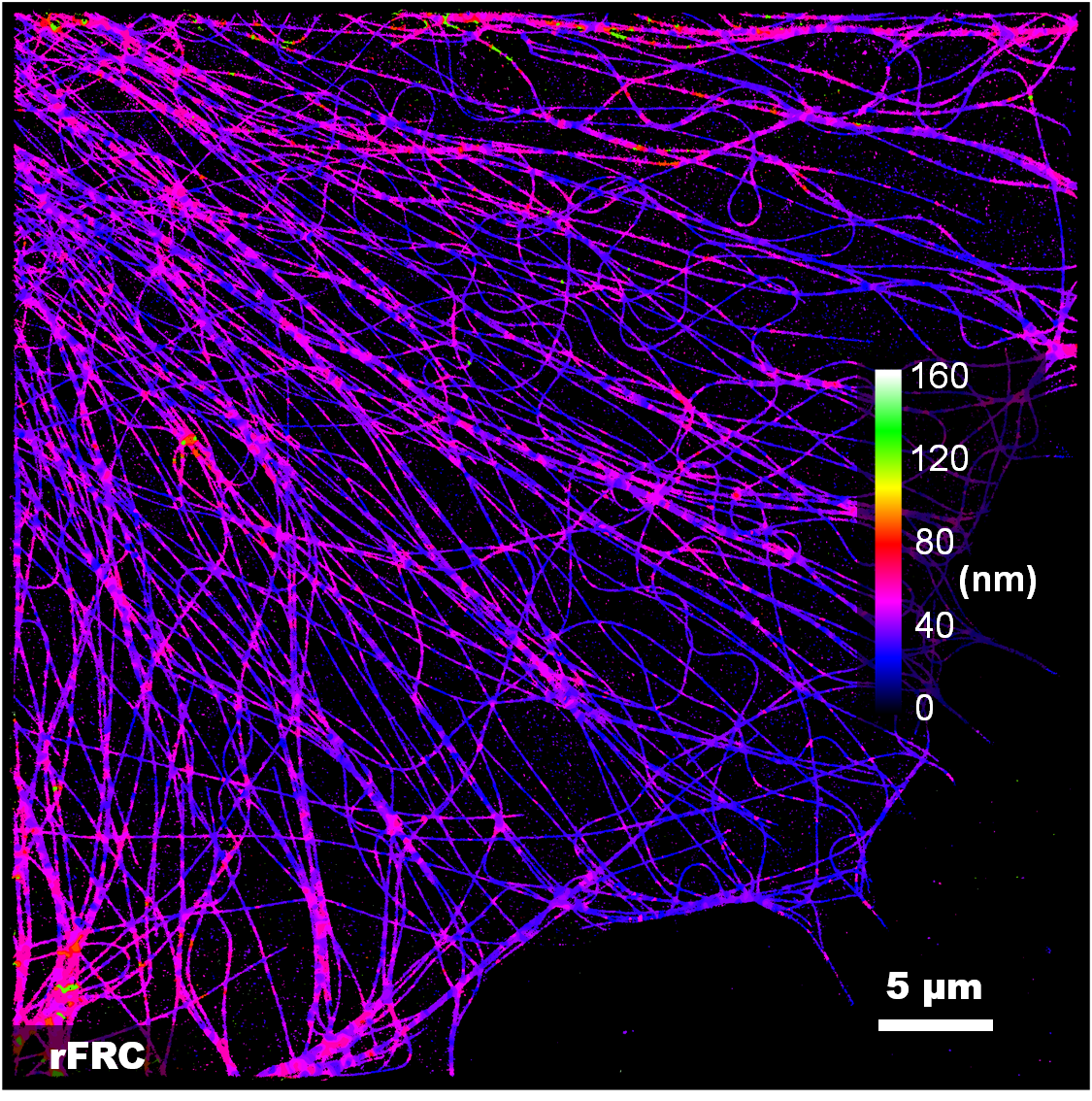
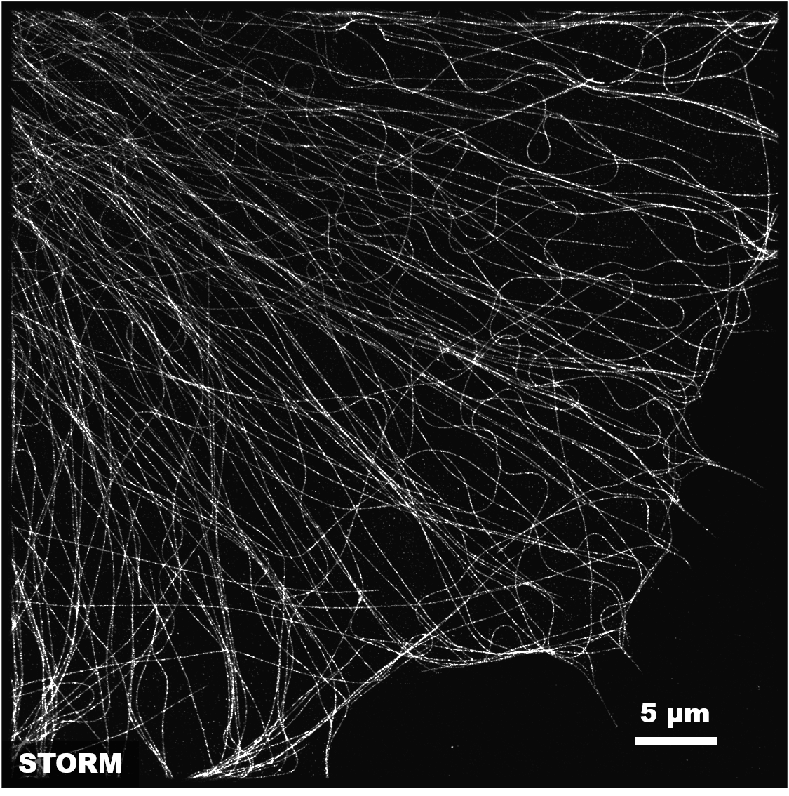
STORM
Objective:
TIRF objective (100×/1.45 oil, Nikon)
Camera:
EMCCD camera (iXon Ultra 897, Andor)
Cell line:
COS-7 cell.
Label:
α-tubulin labeled with Alexa Fluor 647.
Description:
Left: STORM; Right: rFRC resolution map.
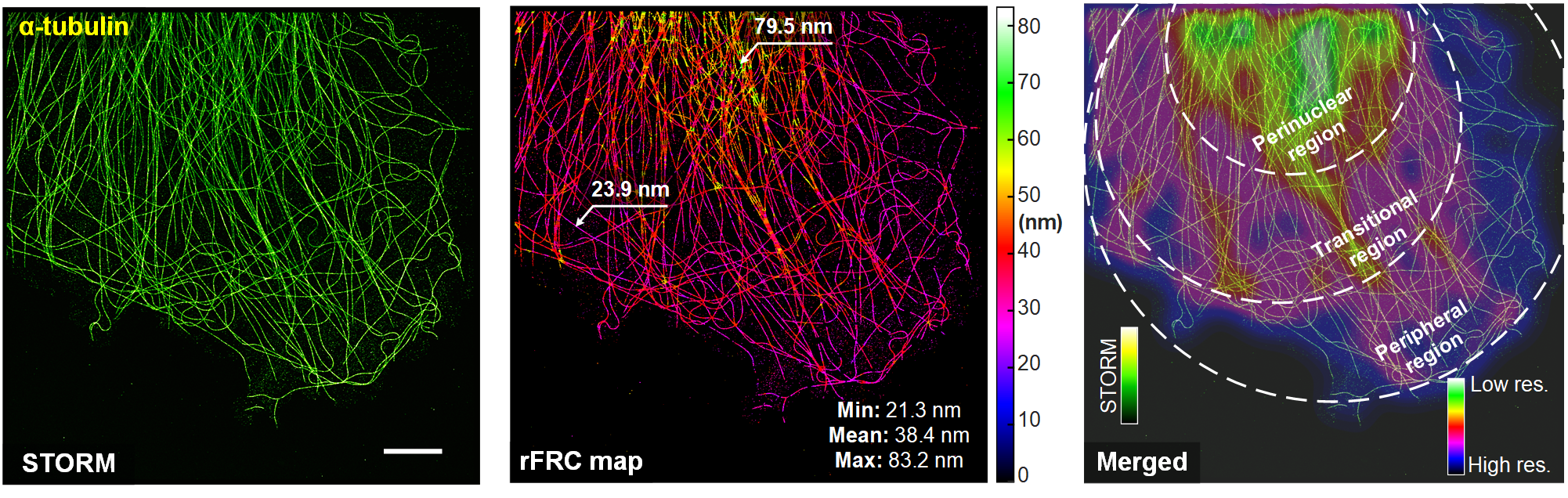
System:
STORM
Objective:
TIRF objective (100×/1.45 oil, Nikon)
Camera:
EMCCD camera (iXon Ultra 897, Andor)
Cell line:
COS-7 cell.
Label:
α-tubulin labeled with Alexa Fluor 647.
Description:
Left: STORM; Right: rFRC resolution map.
MGAN-SIM
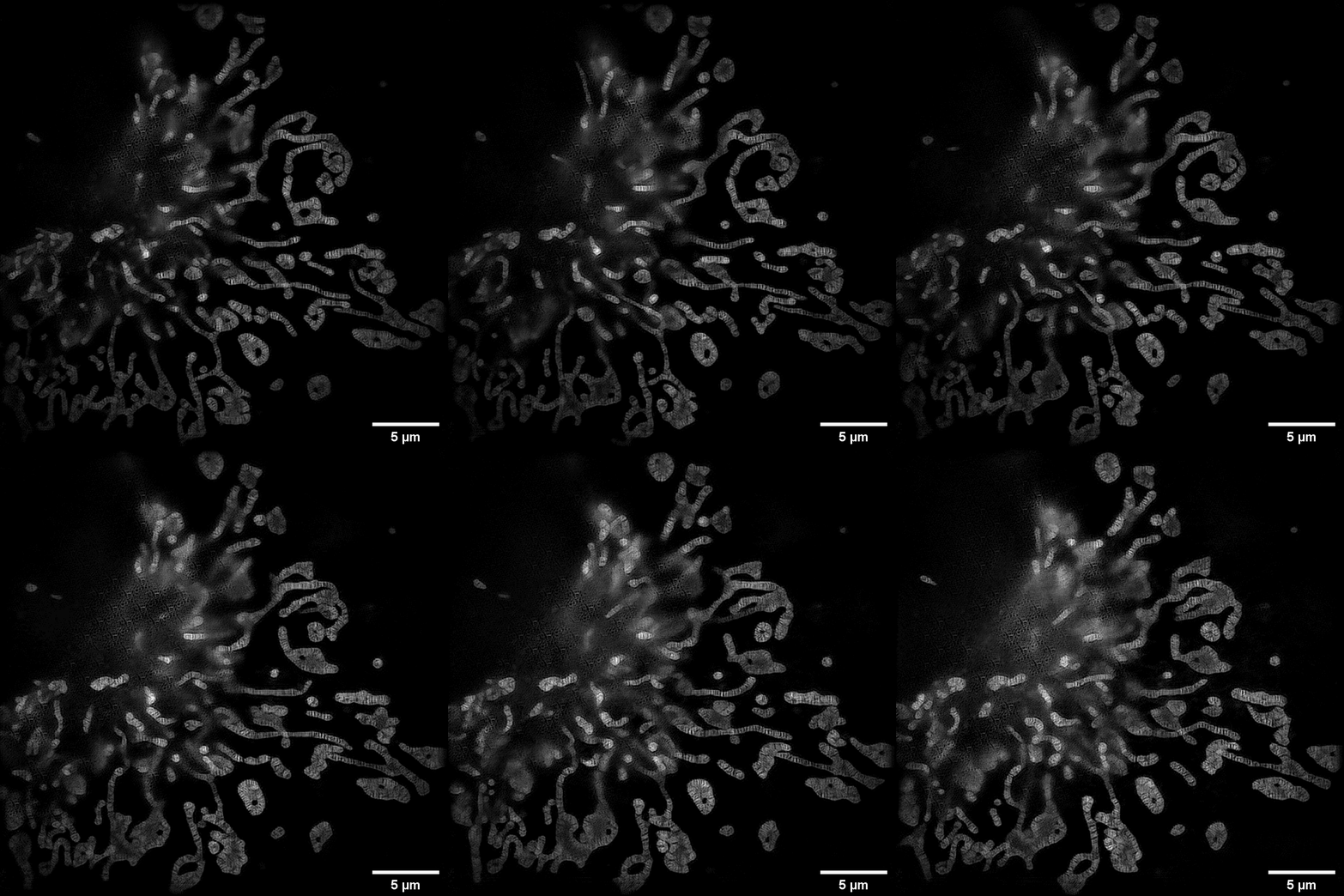
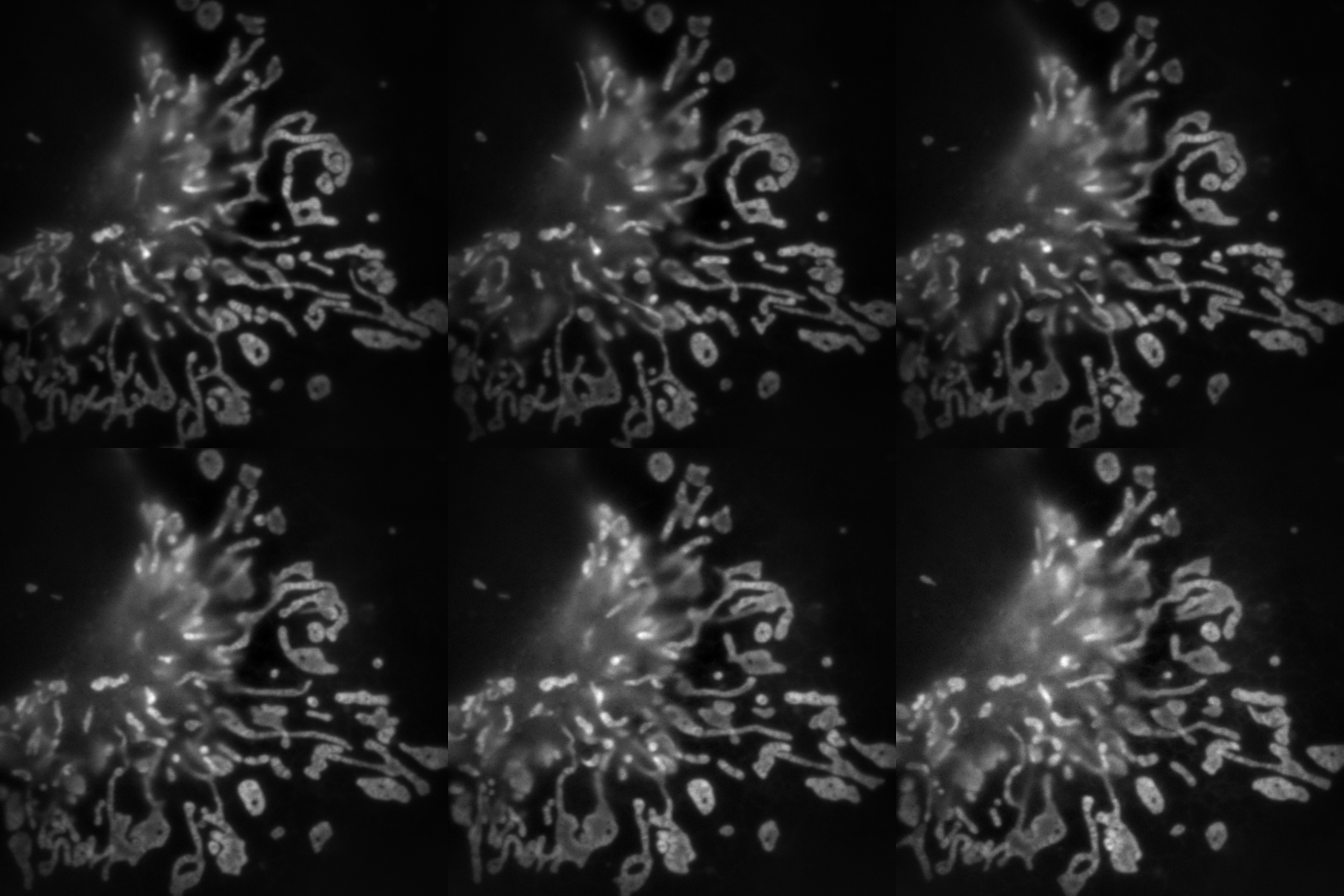
System:
2D-SIM.
Objective:
Wide-field objective (100×/1.45 oil, Olympus)
Camera:
sCMOS camera (Flash 4.0 V3, Hamamatsu, Japan)
Cell line:
COS-7 cell.
Label:
MitoTracker Green.
Description:
Left: wide-field; Right: MGAN-SIM.
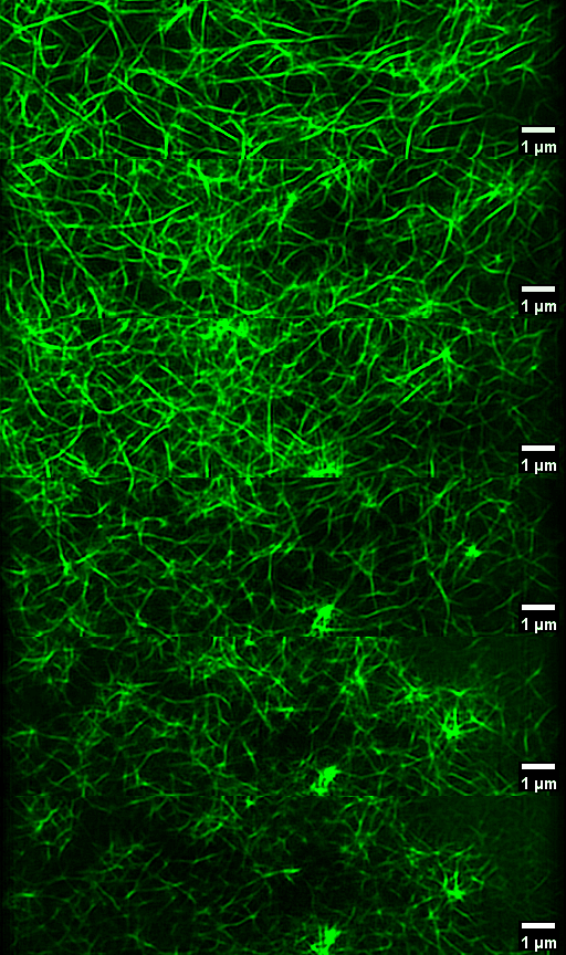
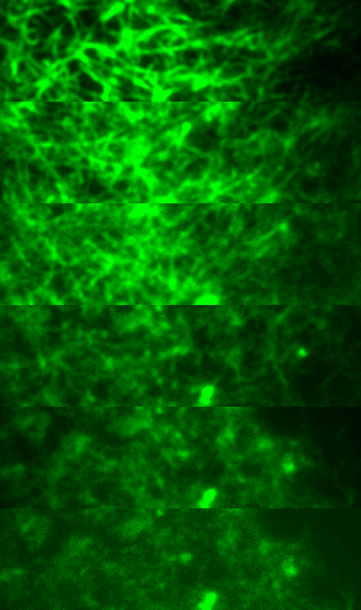
System:
High NA TIRF-SIM.
Objective:
TIRF objective (100×/1.7 HI oil, Olympus)
Camera:
sCMOS camera (Flash 4.0 V3, Hamamatsu, Japan)
Cell line:
INS-1 cell.
Label:
LifeAct-EGFP.
Description:
Left: TIRF; Right: MGAN-SIM.
Long-term 1h imaging, 3400 frames.
ODT
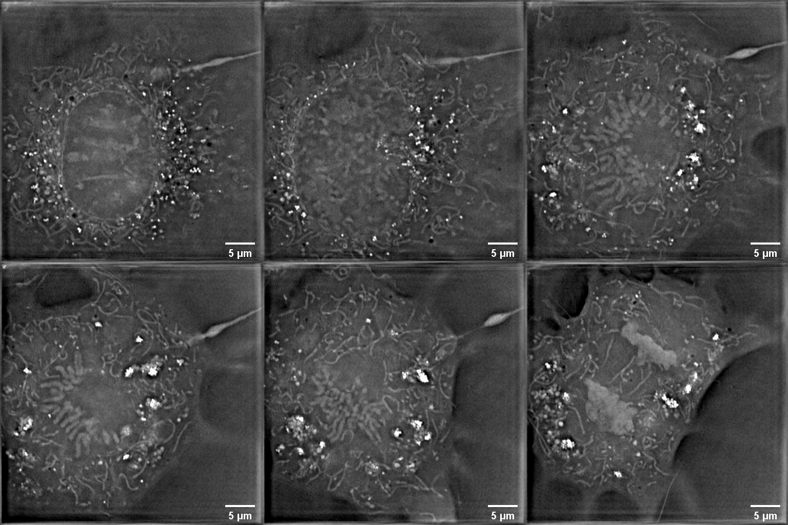
System:
ODT.
Objective:
Wide-field objective (100×/1.49 oil, Olympus)
Camera:
sCMOS camera (Flash 4.0 V3, Hamamatsu, Japan)
Cell line:
COS-7 cell.
Description:
The process of cell division in hours.
0~10 μm and the plane at 7 μm is visualized on the left.
From Prof. Liangyi Chen.
SIM-assisted ODT
Single channel
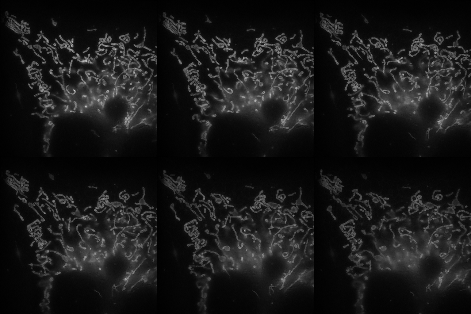
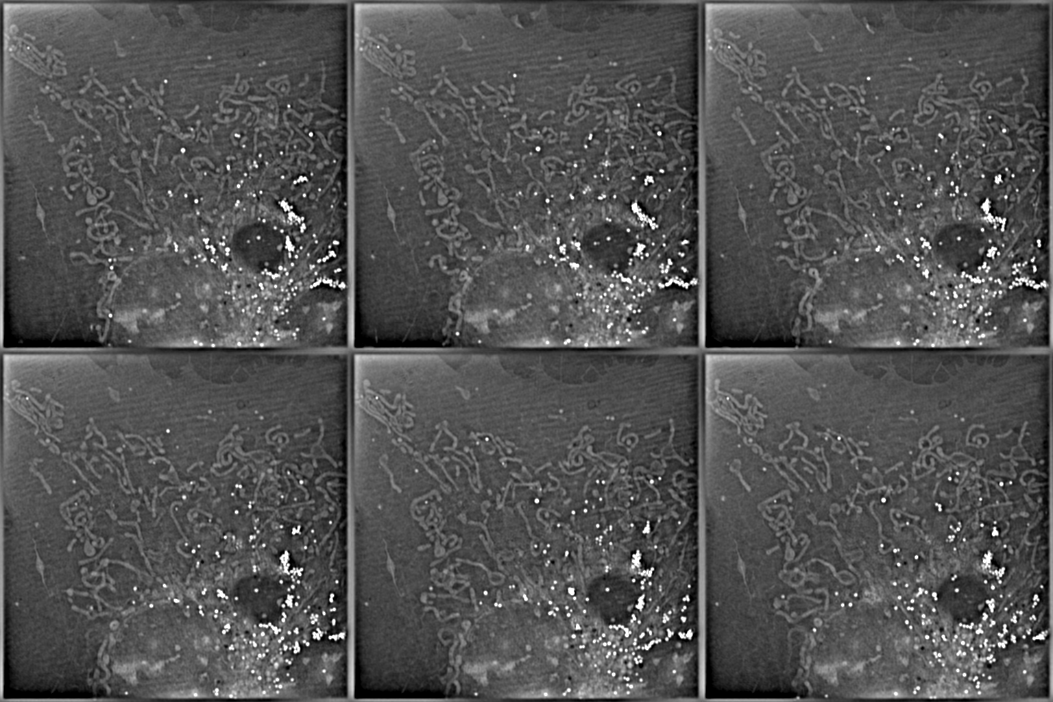
Merged
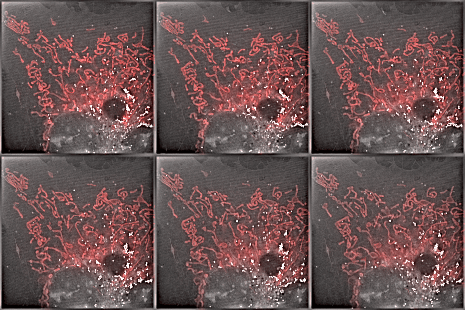
System:
2D-SIM assisted ODT.
Objective:
Wide-field objective (100×/1.49 oil, Olympus)
Camera:
sCMOS camera (Flash 4.0 V3, Hamamatsu, Japan)
Cell line:
COS-7 cell.
Label:
MitoTracker Green.
Description:
Left panel: ODT (left) at 5μm plane (0~6μm in total); 2D-SIM (right).
Right panel: 2D-SIM (red) + ODT (gray).
From Prof. Liangyi Chen.
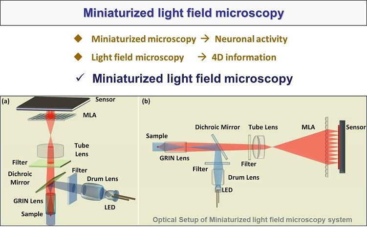
Miniaturized light-field microscopy
System concept
System on a live-mouse
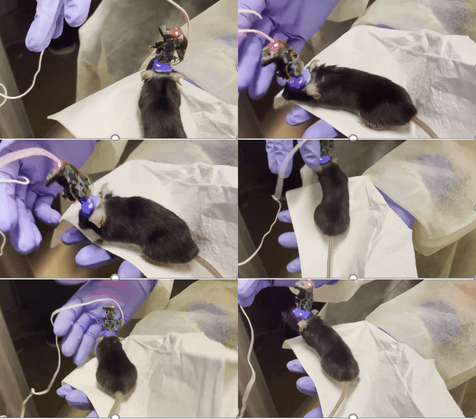
System: Miniaturized light-field microscopy.
Objective: GRIN lens (0.5 NA)
Camera: CMOS camera
Sample: Live-mouse.
Label: GCaMP6s in vivo.
Description: From Changliang Guo.