Projects

Microscopy: Systems & Algorithms

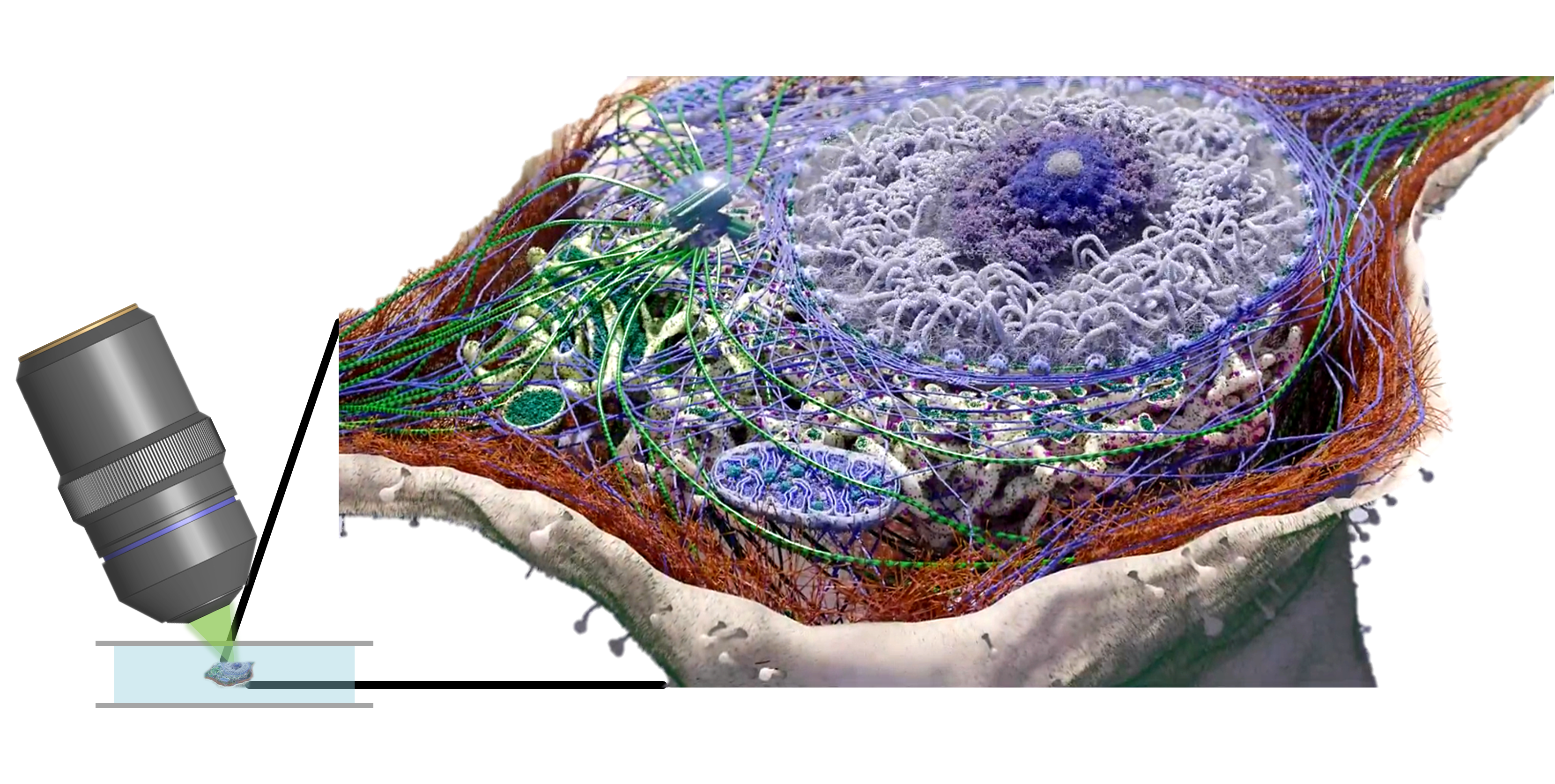
We focus on developing advanced biomedical imaging technologies across modalities. Our research spans new imaging physics, bottom-up system design, and the co-design of hardware and computational algorithms tailored to specific imaging scenarios. We aim to optimize spatial, temporal, and information resolution under practical constraints such as photon or signal budget, acquisition speed, imaging depth, and system complexity.

Representative developed techniques:

(1) Sparse deconvolution is a physics-informed reconstruction framework that exploits sparsity and structural continuity to enhance resolution, contrast, and SNR beyond the limits of microscopy, particularly under low-SNR conditions. Applications include Sparse-SIM and Sparse SD-SIM.

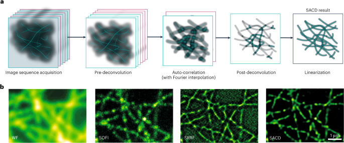
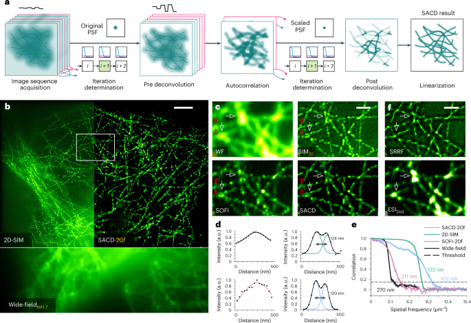
(2) SACD is a fluctuation-based super-resolution framework that achieves approximately threefold lateral and axial resolution enhancement using only 20 frames (10 min for a 2 mm × 1.4 mm FOV), without additional optical components. Sparse-SACD further enables fast 4D live-cell super-resolution imaging. See also our label-free SACD [Light: Science & Applications, 2025].

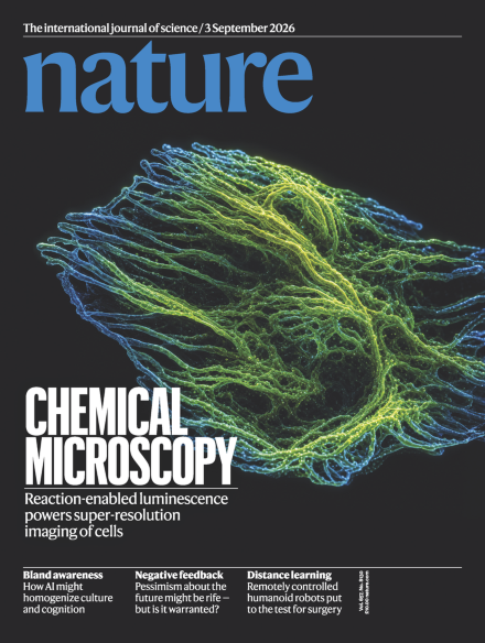
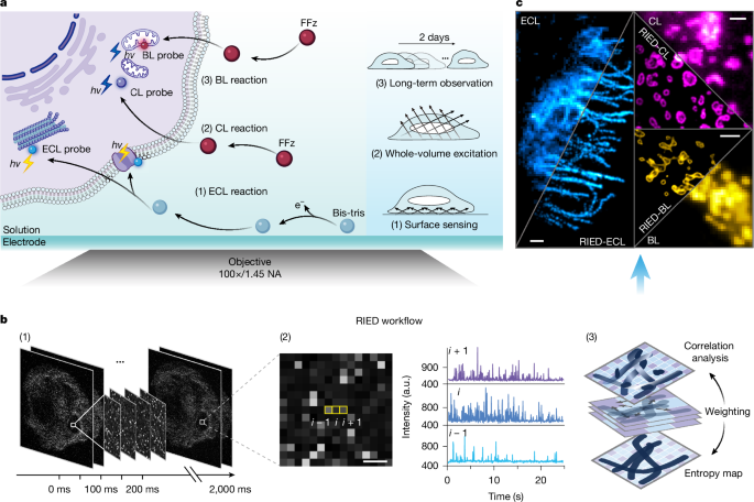
(3) RIED is a reaction-luminescence super-resolution framework for extremely photon-limited bioluminescence (BL) and electrochemiluminescence (ECL). By decoding intrinsic luminescence fluctuations, RIED enables high-contrast 2D and 3D super-resolution imaging without external optical excitation or photochemical modulation. BL-RIED supports continuous super-resolution imaging for over two days.

Microscopy systems and algorithms

From imaging physics and instrumentation to computational reconstruction and system–algorithm co-design.

Machine learning: Methods & Applications

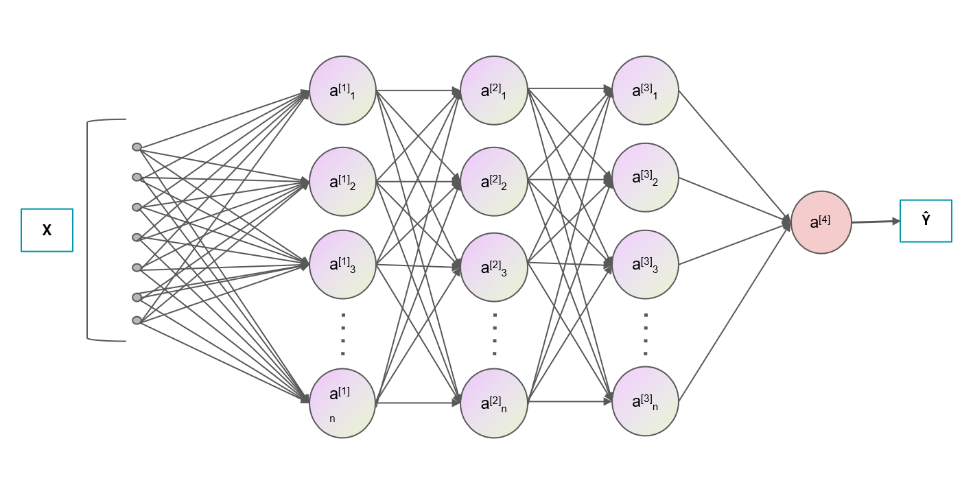
We develop machine-learning methods for the reconstruction and analysis of multidimensional biomedical and natural signals. We exploit both explicit physical and structural priors to design interpretable computational models, and data-driven priors to develop deep-learning approaches for problems that are difficult to address with conventional methods. Our work includes self-supervised learning, representation learning, Bayesian learning, and foundation models, with applications ranging from reducing the photon, acquisition-time, and hardware requirements of microscopy to accelerating biological image reconstruction, profiling, and discovery.

Representative developed techniques:

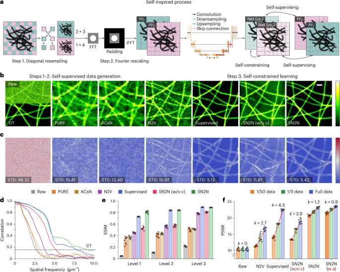
(1) SN2N (Self-inspired Noise2Noise) is a self-supervised learning-to-denoise framework that achieves performance competitive with supervised learning while eliminating the need for large paired datasets and clean ground-truth images. It enables robust denoising across diverse biomedical imaging modalities and supports photon-efficient, long-term, and low-phototoxicity imaging.

(2) aSN2N (adaptive SN2N) introduces adaptive preprocessing and normalization strategies to stabilize self-supervised learning across heterogeneous imaging conditions. It improves robustness to intensity variations and background contamination while effectively suppressing reconstruction artifacts.

Machine learning for biomedical imaging

From physical and structural priors to self-supervised, representation, and foundation-model learning.

Smart Bioimage Analysis & Biomedical Devices

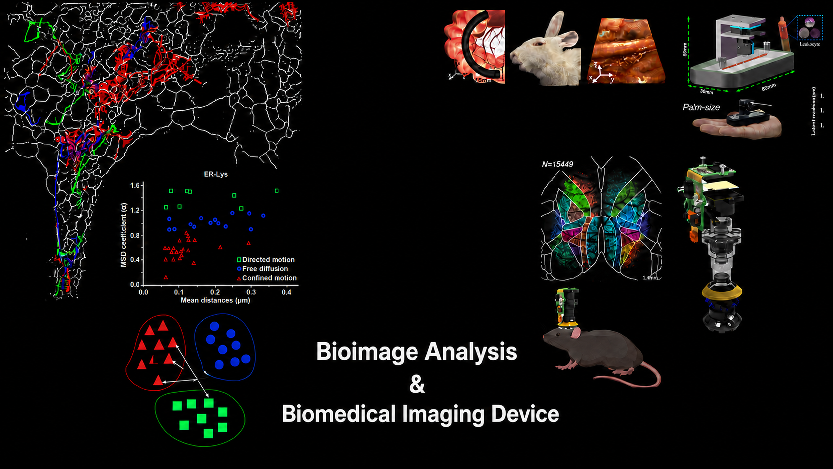
We develop intelligent bioimage analysis methods and biomedical imaging instrumentation to enable automated, quantitative, and high-throughput biological investigation. Our work spans image segmentation, tracking, profiling, and representation learning, as well as the development of compact, application-oriented imaging devices and systems.

Representative developed techniques:

(1) PANEL (pixel-level analysis of error locations) is a quantitative framework for mapping reconstruction errors at the super-resolution scale, enabling systematic assessment of both conventional and deep-learning-based reconstruction methods.

(2) Smart palm-size optofluidic hematology analyzer is a compact imaging-based platform for automated leukocyte concentration measurement and rapid hematological analysis.

(3) Light-field endoscopic probes initiated with MIRD (multiple micro-imaging devices), provide highly flexible and compact probes for single-shot, high-resolution volumetric imaging. We continue to develop new optical designs and computational imaging strategies to improve imaging performance, probe flexibility, and practical applicability. See also [Optics and Lasers in Engineering, 2026] and [Optics Express, 2026].

(4) Sparse confocal microscopy with single-particle tracking was applied to reveal how SARS-CoV-2 virus-like particles exploit filopodia to reach cellular entry sites through two distinct modes, “surfing” and “grabbing”, thereby reducing random searching on the plasma membrane. See also [European Journal of Cell Biology, 2025].

Smart bioimage analysis and biomedical devices

From quantitative bioimage analysis to compact and application-oriented biomedical devices.

News from the Lab

2026-08: Our RIED work is online at Nature; RIED paper preview X · August 12, 2026 RIED is out at Nature A new paradigm for excitation-free super-resolution microscopy using ECL, CL, and BL. View post on X ↗ 2026-07: Congrats to Jiahui Gui, the Ph.D. student from the lab and co-first author, on the acceptance of our RIED work in Nature!

2026-04: Congrats to Deer Su, the Ph.D. student from the lab, on successfully defending his Ph.D. dissertation!

2026-04: Congrats to Deer Su, the Ph.D. student from the lab. Two studies advancing light-field endoscopic probes have been published in Optics and Lasers in Engineering and Optics Express;

2026-02: Congrats to Jiahui Gui, the Ph.D. student from the lab. RIED is released as a bioRxiv preprint, together with the MATLAB toolkit;
2025-09: The lab PI has been promoted to Tenured Professor at Harbin Institute of Technology;

2025-09: Congrats to Liying Qu, the Associate Investigator from the lab. Our commentary on SN2N is online at Clinical and Translational Medicine;

2025-08: FLAME is released as a preprint, together with its MATLAB toolkit;

2025-08: Our collaborative work, EPSLON, has been published in Light: Science & Applications;

2025-07: SR-Wiki is now the lab’s official GitHub organization for releasing our latest imaging and analysis tools and provides implementations of the lab’s imaging toolkit.

2025-06: Congrats to Jingyang Zhu, the Ph.D. student from the lab. Adaptive SN2N has been published in PhotoniX Life;

2025-04: Congrats to Liying Qu on successfully defending her Ph.D. dissertation;
2024-09: Our SN2N work is online at Nature Methods; SN2N paper preview X · September 11, 2024 SN2N is out at Nature Methods An unsupervised denoising solution competitive with supervised learning, trained from a single noisy frame without clean ground truth. View post on X ↗ 2024-08: The lab PI has received support from the Excellent Young Scientists Fund of the National Natural Science Foundation of China;

2024-06: Congrats to Liying Qu & Yuanyuan Huang, the Ph.D. students from the lab. SN2N has been accepted by Nature Methods for publication;

2024-01: Congrats to Liying Qu & Yuanyuan Huang, the Ph.D. students from the lab. SN2N is released as a bioRxiv preprint (under review at Nature Methods), together with its Python toolkit;

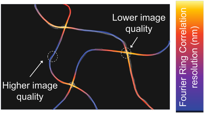
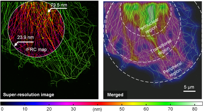
2024-01: Dr. Ke Xu wrote a News & Views in Light: Science & Applications entitled “Mapping super-resolution image quality” for our rFRC work; Mapping super-resolution image quality paper preview Light: Science & Applications · News & ViewsMapping super-resolution image qualityA News & Views article by Ke Xu discussing our rFRC work for local super-resolution image-quality mapping.Read the article ↗ 2024-01: The lab PI has been appointed as a Full Professor;

2024-01: The lab PI now serves as an Associate Editor at npj Imaging; npj Imaging editorial board preview npj Imaging · Editorial BoardAssociate Editor at npj ImagingThe lab PI now serves as an Associate Editor at npj Imaging.View the editorial board ↗
2023-12: The rFRC work is online at Light: Science & Applications; rFRC paper preview X · December 14, 2023 rFRC is out at Light: Science & Applications A tool for evaluating super-resolution image quality at the corresponding super-resolution scale. View post on X ↗ 2023-12: Post a 'behind the paper' blog regarding the rFRC at Nature Research Community;

2023-12: Congrats to Deer Su, the Ph.D. student from the lab. The Smart palm-size optofluidic hematology analyzer is online at Opto-Electronic Science (invited);

2023-12: Congrats to Deer Su, the Ph.D. student from the lab. The MIRD volumetric endoscope is online at Optics Letters, and is selected as the featured image of VOLUME 48, ISSUE 24;

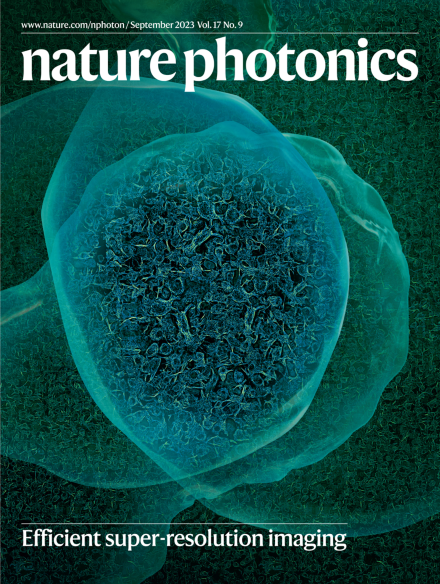
2023-09: Dr. David Baddeley wrote a News & Views article, “Deconvolution enhances fluctuation detection,” in Nature Photonics for our SACD work; Deconvolution enhances fluctuation detection paper preview Nature Photonics · News & ViewsDeconvolution enhances fluctuation detectionA News & Views article by David Baddeley discussing our SACD work and its two-step deconvolution workflow.Read the article ↗ 2023-09: The SACD is selected as the cover article of Nature Photonics Volume 17 Issue 9, September 2023;

2023-08: Using Sparse deconvolution-enhanced confocal microscopy, we (with Yaming Jiu's group) reveal the processes of SARS-CoV-2 regulating and utilizing dynamic filopodia for viral invasion, published online at Science Bulletin;

2023-06: Post a 'behind the paper' blog regarding the SACD at Nature Research Community;

2023-06: The SACD is online at Nature Photonics; SACD paper preview X · June 15, 2023 SACD is out at Nature Photonics SACD enhances fluorescence-fluctuation detection sensitivity by orders of magnitude and needs only 20 frames for >2-fold 3D resolution. View post on X ↗
2022-12: PANEL and SACD are released as preprints;

2022-10: PANELpy is fully open-source;

2022-07: Joined Harbin Institute of Technology as an Assistant Professor;

2022-05: SACDm and SACDj are fully open-source;

2021-11: A Python version with GPU acceleration of the Sparse deconvolution is released at GitHub;

2021-11: Post a 'behind the paper' blog about the Sparse deconvolution at Nature Research Community;

2021-11: The Sparse deconvolution is online at Nature Biotechnology;

2021-07: PANELM and PANELJ are fully open-source;

2021-07: The Sparse deconvolution is accepted by Nature Biotechnology for publication;

2021-03: An OPEN scientific discussion about deconvolution is posted on GitHub as well as on Twitter;